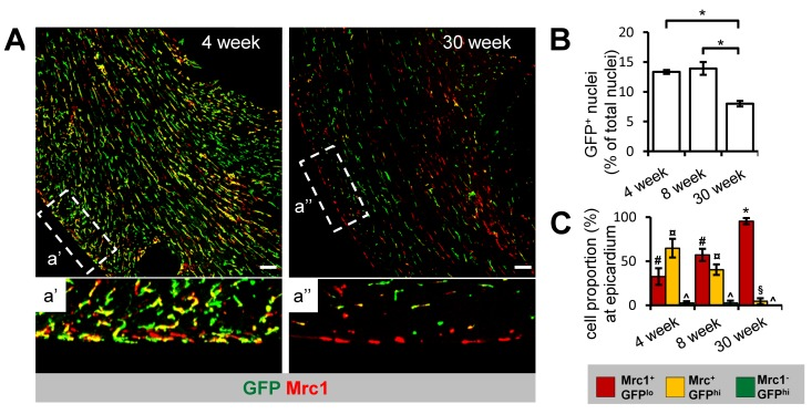
Figure 3.Age-dependent decline in Cx3cr1+ cTM cell density and proportion at the epicardium(A) 45 μm confocal micrograph maximum intensity view of GFP and Mrc1 staining in mouse hearts sections from 4 and 30 week old Cx3cr1GFP/+ mouse heart sections (scale bar indicates 100 μm). a' and a'' are magnified views of areas demarked by dotted lines. (B) Histogram of proportion of GFP+ nuclei in relation to total nuclei in hearts of 4, 8 and 30 week old Cx3cr1GFP/+mouse heart sections. *p≤0.05. (C) Histogram of proportion of Mrc1+GFPlo, Mrc1+GFPhi or Mrc1−GFPhi cTMs in relation to all cTMs at the epicardium. #p>0.05 Mrc1+GFPhi vs Mrc1+GFPlo; ¤p≤0.05 Mrc1−GFPhi vs Mrc1+GFPhi; ^p≤0.05 Mrc1−GFPhi vs Mrc1+GFPlo; *p≤0.05 Mrc1+GFPhi vs Mrc1+GFPlo; p>0.05 Mrc1−GFPhi vs Mrc1+GFPhi. All histograms show means ± SEM. Cell numbers were quantified from at least 4 fields of view from 3 mouse hearts/age group