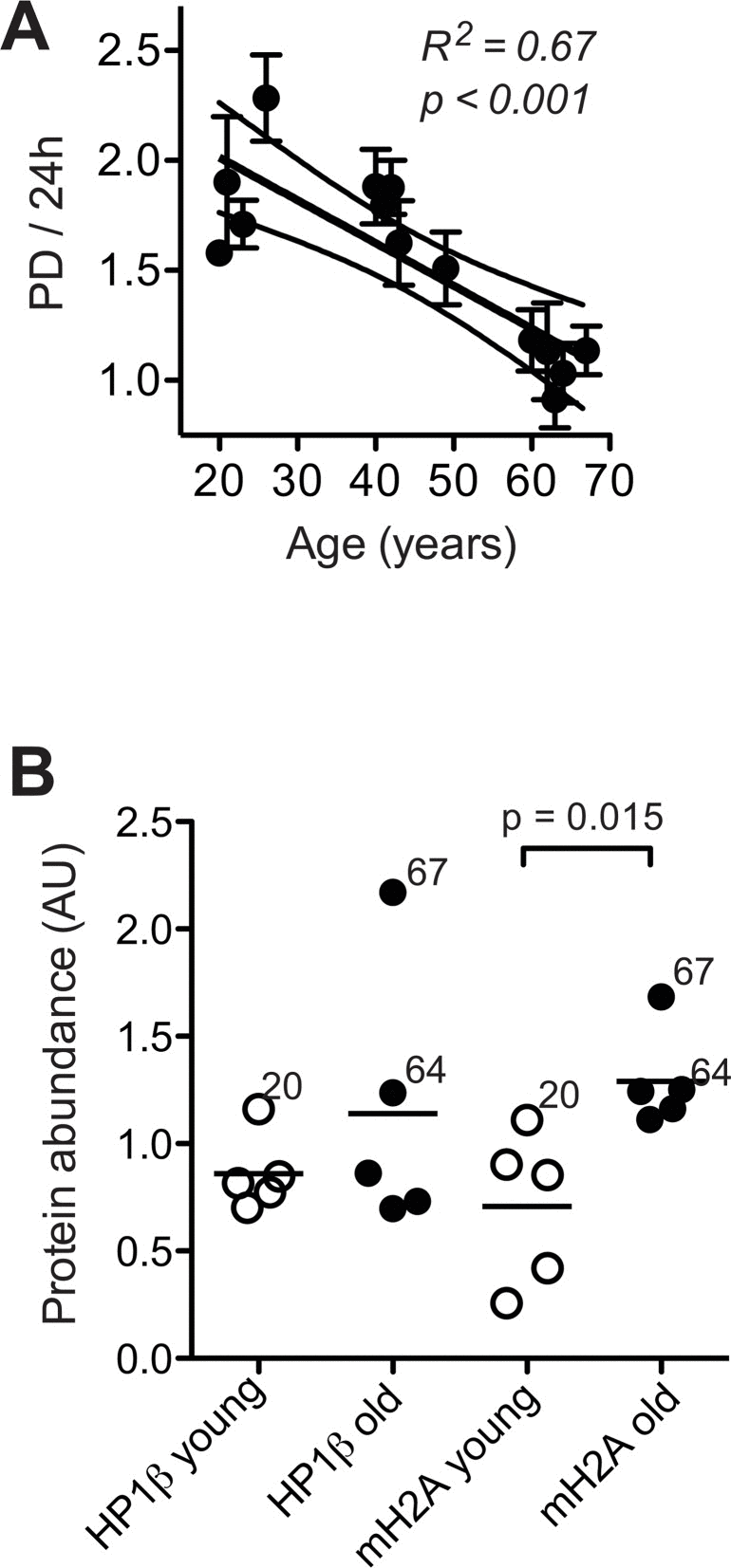
Figure 2.Cell proliferation and heterochromatin marks(A) Mean ± SEM of population doublings (PD) per 24 h determined for each donor in five to six independent cultures by seeding a defined amount of cells onto a standardised area of substratum and monitoring the time required for growth to confluency and the final cell yield. Results of linear regression are stated as R2: Pearson's coefficient for goodness of fit, p: probability for slope = 0, dotted lines: 95% confidence limits of linear regression, and N.S.: linear regression of the data revealed no significant age-related change in the parameters. (B) Heterochromatin marks HP1β (left) and mH2A (right) determined by immunoblotting in whole cell lysates of dermal fibroblasts from the young (open symbols) and old donor group (closed symbols) at PD < 14. Data were normalised to values obtained in control fibroblasts subjected to replicative senescence. Data points represent means of triplicate determinations in separate cell cultures. Errors were < 30% of the values and are omitted for the sake of clarity. Horizontal lines indicate the mean of the respective age group. Numbers next to data points indicate the chronological age of the respective donor.