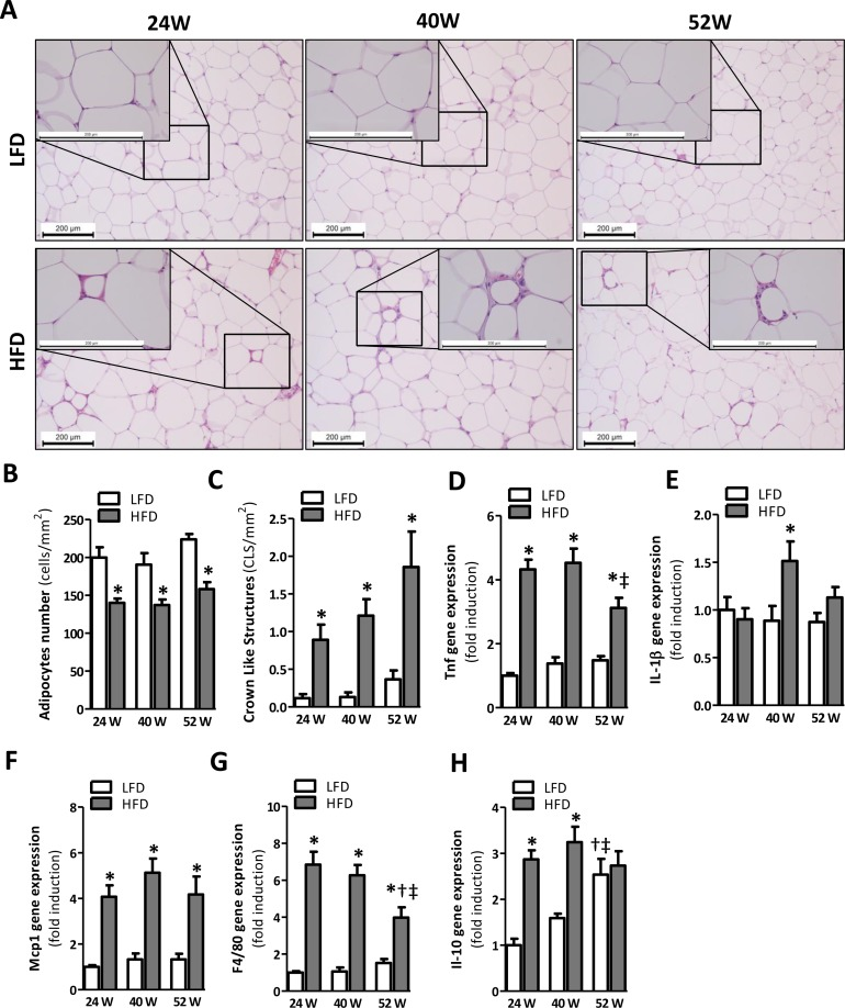
Figure 3.Prolonged HFD-feeding leads to AT inflammation in mice after 24 weeks(A) Representative pictures from H&E-stained AT sections of LFD and HFD mice after 24 (left), 40 (middle) and 52 (right) weeks of diet with crown-like structures (insets). Histologically quantified number of (B) adipocytes and (C) crown-like structures per mm2 (n=10-15). (D-H) mRNA expression levels of tumor necrosis factor (Tnf), interleukin-1β (Il1β), monocyte chemotactic protein-1 (Mcp1), macrophage marker (F4/80) and interleukin-10 (Il-10) in the AT. All mRNA expression data were normalized to the LFD24 group and expressed as mean ± SEM (n=7-8). Significance level set at p<0.05. *=significant from LFD at same time point, †=significant from same diet 24w, ‡=significant from same diet 40w.