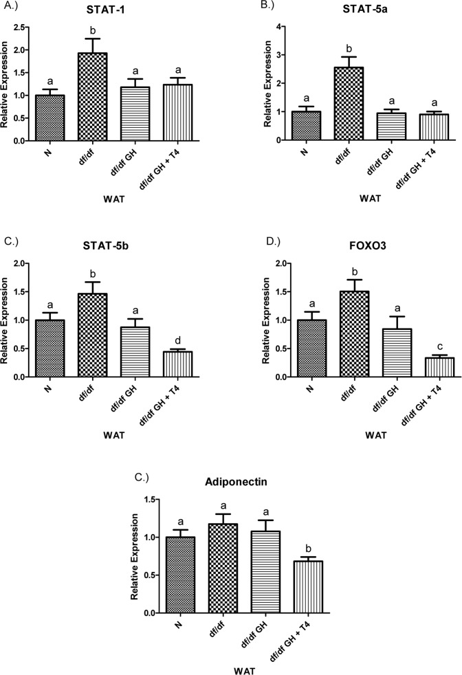
Figure 9.(A) Signal Transducer and Activator of Transcription (STAT) ‐1, (B) STAT‐5a, (C) STAT‐5b, (D) Adiponectin, and (E) FOXO3 relative gene expression in female Ames dwarf (df/df) mouse white adipose tissue after growth hormone (GH) and thyroxine (T4) treatment. For A‐D, wild‐type (N)n=10;df/dfn =9;df/dfGHn= 9;anddf/dfGH+T4n=8. ForE(FOXO3), Normal(N) n = 10;df/df n=9;df/dfGHn= 9;anddf/df GH+ T4n= 7. Groups that do not share a superscript show differences with statistical significance (p < 0.05). Both GH and GH + T4 treatment decreased PPAR-γ gene expression level in WAT of df/df mice (compared with df/df mice, p <0.05, p< 0.005 respectively). Moreover, animals in the df/df-GH + T4group had lower PPAR-γ expression than the df/df-GH group (p < 0.05) (Figure 8), PGC-1α mRNA levels were significantly increased in the WAT of Ames dwarf mice compared with N controls (p < 0.005).
Figure 9 — Thyroxine modifies the effects of growth hormone in Ames dwarf mice | Aging