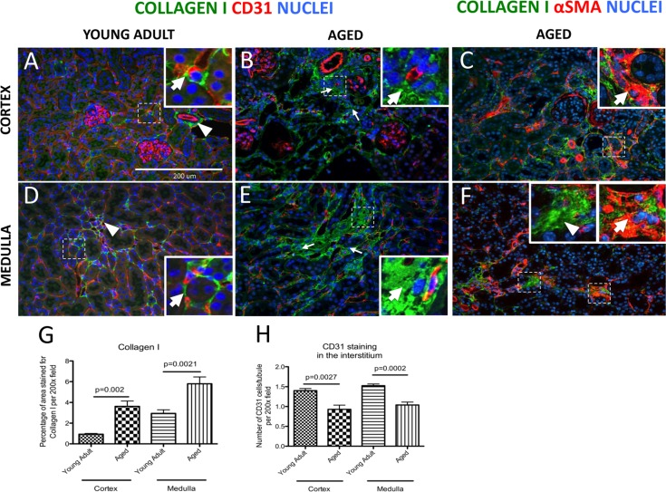
Figure 1.Increase in interstitial fibrosis and reduction of microvascular density in aged mice kidneysFibrosis was measured by staining for collagen I (green color). Double staining of αSMA (intracellular antigen) and collagen I was used to discern collagen I-expressing myofibroblasts from extracellular matrix proteins. Microvascular rarefaction was assessed by CD31 staining (red). DAPI staining identifies nuclei (blue). (A) Collagen I staining was faint and confined to the fibroblasts/pericytes in the cortex of young adult kidneys. The inset shows a high power image of a collagen I+ cell (green) encircling capillary (red) (arrow). Adventitial cells of renal arteries also stain for collagen I (arrowhead). (B) Collagen I staining was more abundant in the cortical interstitium of aged kidneys (arrows indicate examples) and was found outside the capillary walls (inset, arrow shows collagen I staining). CD31 staining was reduced in intensity. (C) Co-staining for collagen I and αSMA shows the presence of myofibroblasts (inset, arrow). (D) Collagen I staining in the medulla of young adult mice was present in vasa recta (arrowhead) and perivascular fibroblasts (inset, arrow). (E) In aged kidneys interstitial collagen I staining was increased (arrows indicate examples). The inset shows high power view of accumulation of collagen I staining (arrow). CD31 staining was decreased. (F) Co-staining of collagen I with αSMA confirms the presence of myofibroblasts (inset, arrow). Extracellular collagen I was also detected (inset, arrowhead). (G) Graph showing quantification of interstitial collagen I staining was significantly increased in both the cortex and the medulla. (H) Quantification of CD31 staining shows reduced vascular density per tubule in both the cortex and the medulla. Data are represented as mean ± SEM (n=6).