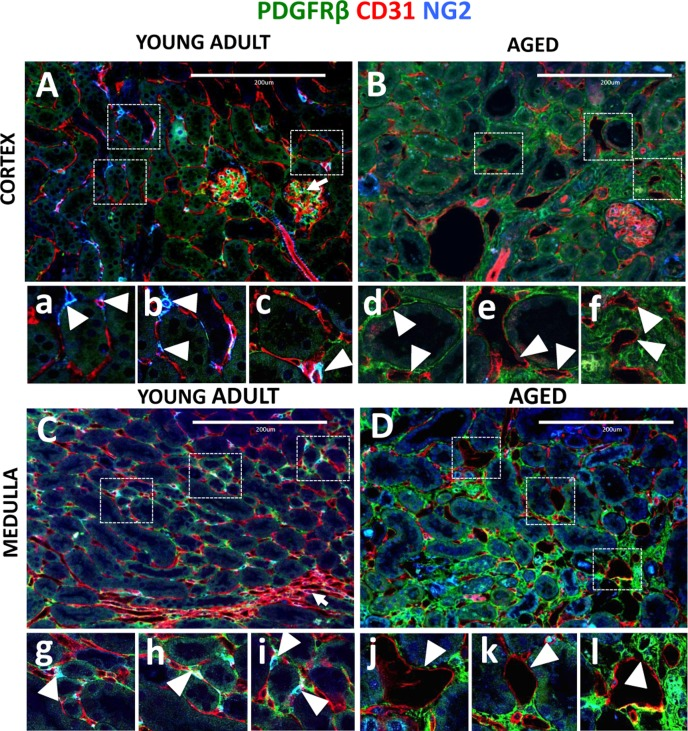
Figure 4.Capillary dilation in aged mice was associated with reduced pericyte coverageEndothelial cells were marked by CD31 expression (red color). Pericytes were labelled by NG2+/PDGFRß+ staining (blue and green colors respectively). Double positive cells (light blue) were quantified using single channel images. (A) In young adult kidney cortex, peritubular capillaries form a regular pattern around renal tubules and inside the glomerular tuft (arrow). The boxed regions show higher power images of interstitial pericytes supporting peritubular capillaries (a, b, c, arrowheads). (B) In aged kidney cortex, endothelial cells become dilated, have decreased pericyte coverage (d, e, arrowheads) and some were completely isolated from tubules (f, arrowheads). (C) In young adult medulla, endothelial cells occupy peritubular spaces and vasa recta (arrow). Typically, few capillaries surround each tubule and pericyte, and are closely attached to the endothelial cells (g, h, i, arrowheads). (D) In aged kidney medulla, dilated peritubular capillaries were also present (j, k, arrowheads). In some instances, dilated capillaries appear in the areas of increased interstitial PDGFRß+ staining (l, arrowhead).