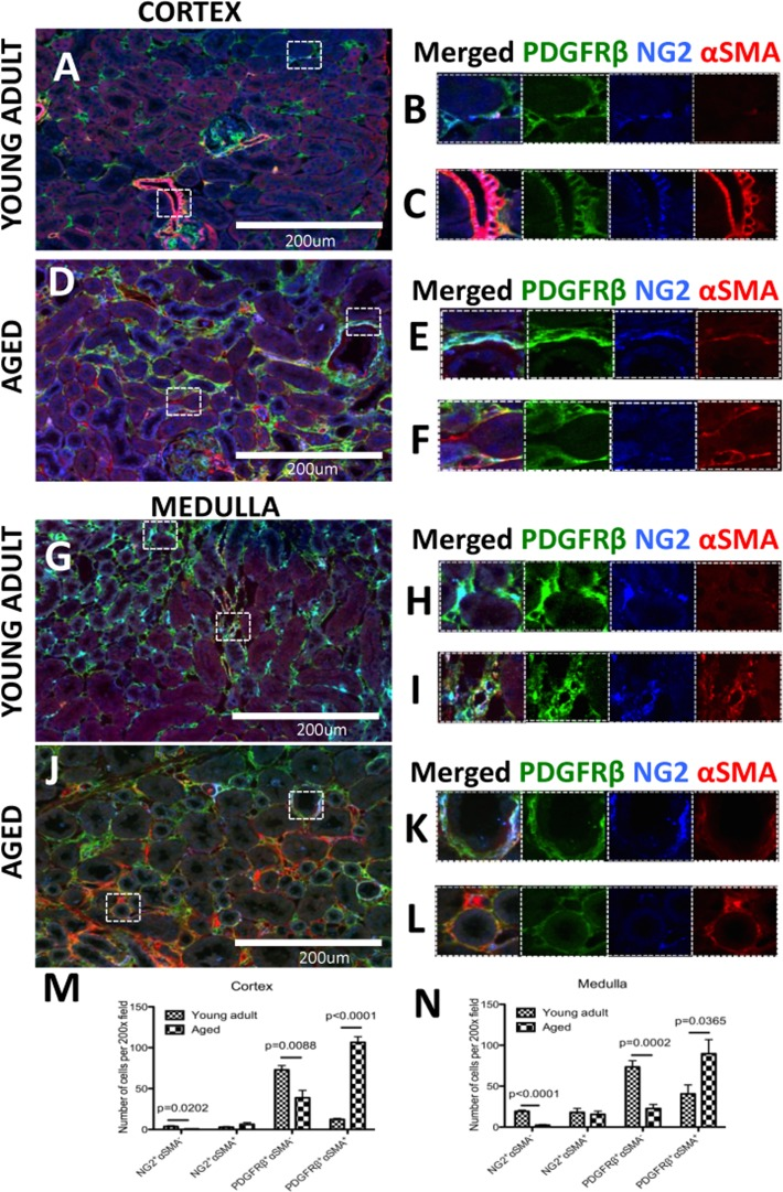
Figure 5.A subset of pericytes differentiate into myofibroblasts and increase in aged kidneysPericytes were identified by NG2+/PDGFRß+ staining (blue and green colors respectively). αSMA was used as myofibroblast marker (red color). (A) In the young adult kidney cortex, (B) most pericytes do not express αSMA. However in preglomerular arterioles (C) αSMA expression was present together with NG2/PDGFRß staining. (D) In aged kidney, αSMA increased in pericytes (E) and PDGFRß+ /NG2− cells (F). (G) Medulla of young adult kidney showed the presence αSMA in some peritubular capillaries (H) and contractile vasa recta cells (I). In aged mouse kidney, (J) there was accumulation of αSMA+ cells co-expressing PDGFRß and NG2 (K) or PDGFRß only (L). Quantification of αSMA expression in NG2+ and PDGFRß + cells showed that PDGFRß+ cells outnumber NG2+ cells in both young adult and aged kidneys. There was a dramatic increase in PDGFRß+αSMA+ cells and the numbers of αSMA-cells expressing either PDGFRß or NG2 were significantly lower in both (M) the cortex and (N) the medulla. Data are represented as mean ± SEM (n=6).