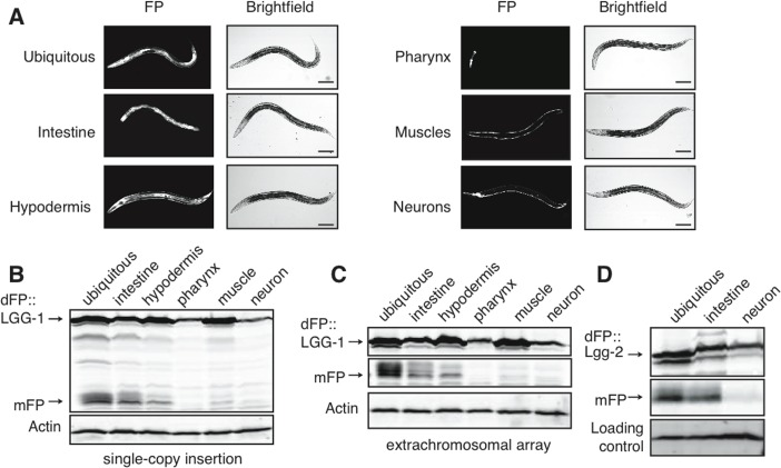
Figure 2.Tissue-specific expression of dFP::LGG-1 reveals variance in basal autophagy(A) C. elegans strains were created with dFP::LGG-1 expressed under control of a promoter that is ubiquitously active (eft-3p), or under the control of promoters driving expression specifically in the intestine (vha-6p), hypodermis (dpy-7p), muscles (myo-3p), pharynx (myo-2p), or neurons (rab-3p). An image of each strain shows fluorescence consistent with the expected tissue-specific expression. Animals were imaged at approximately late L3 larval stage; scale bar is 100mm. Visualizing the fluorescent protein using an anti-FP immunoblot revealed expression in mixed-age populations of strains with the reporter present in either (B) single-copy chromosomal insertion or (C) tandem, multi-copy extrachromosomal array. In both expression conditions mFP is most abundant in the intestine with some mFP present in the hypodermis. A full blot is shown in (B) to provide context, while subsequent images focus on the upper and lower bands of interest; uncropped blots are shown in Supplementary Figure 1. (D) dFP::LGG-2 expressed from the same ubiquitous, intestinal, or neuronal promoter showed lysosomal processing primarily in the intestines.