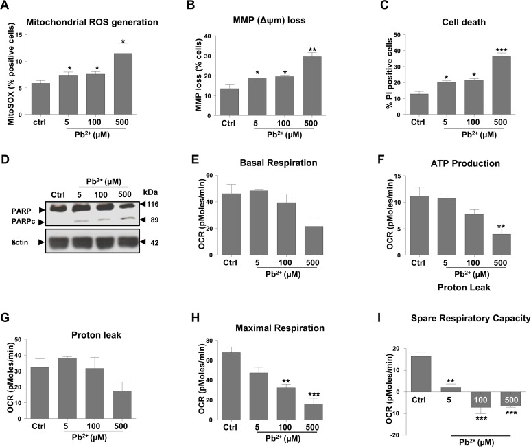
Figure 1.Exposure to lead induces mitochondrial dysfunction and subsequent apoptosis of N27 dopaminergic neuronsCells were incubated for 48-hours with lead acetate, at the concentrations shown. (A) Mitochondrial superoxide levels were measured using MitoSOX staining followed by FACS analysis. (B) The effect of lead treatment on mitochondrial membrane potential (MMP) loss was detected by DiOC6(3). (C) Analysis of cell death measured by propidium iodide (PI). (D) Western blot analysis of PARP cleavage (PARP to PARPc) following lead exposure. Profiles of different parameters of mitochondrial function (E-I) were determined using Seahorse XF24 Analyser. *P<0.05, **P<0.01, ***P<0.001, n=3; mean ± SE.