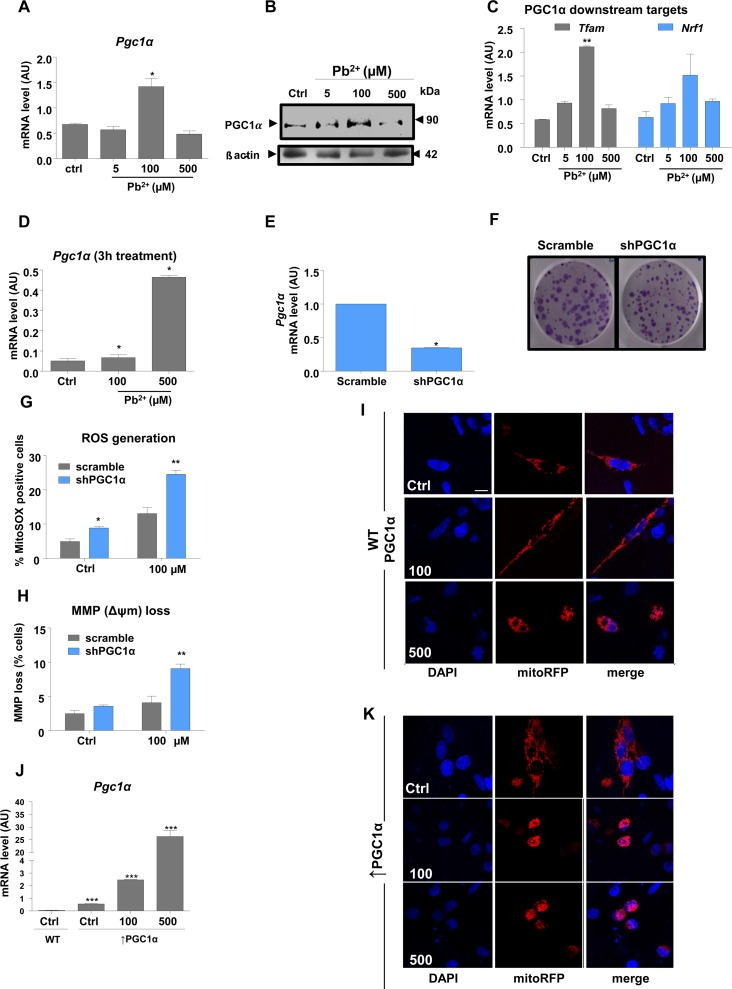
Figure 2.PGC1α protects N27 dopaminergic neuronal cells from Pb 2+ -induced neurotoxicityCells were incubated with lead acetate at the concentrations shown. (A,B) qRT-PCR and western blotting measurement of mRNA and protein expression levels, respectively of Pgc1α in N27 cells treated with lead for 48-hours. (C) mRNA levels of Pgc1α target genes (Tfam, Nrf1)inN27 cells treated with lead for 48-hours.(D)N27 Pgc1αmRNA levels after 3-hours lead treatment.(E) Pgc1α mRNA levels and (F) colony formation in N27 cells with stable downregulation of PGC1α. (G) Mitochondrial superoxide levels were measured in shPGC1α cells using MitoSOX staining and (H) the effect of lead treatment on mitochondrial membrane potential loss in shPGC1α was detected by DiOC6(3) in N27 cells treated with lead for 48-hours. (I,K) Confocal imaging analysis of mitochondrial morphology in wild type PGC1α and in overexpressed PGC1α N27 cells ((Pgc1α) was investigated using Mito Tracker and DAPI staining recorded by fluorescence microscopy. (J) qRT-PCR analysis of Pgc1α in N27 cells expressing exogenous Pgc1α versus(Pgc1α cells, treated with lead for 48-hours.*P<0.05, **P<0.01, n=3; mean ± SE.