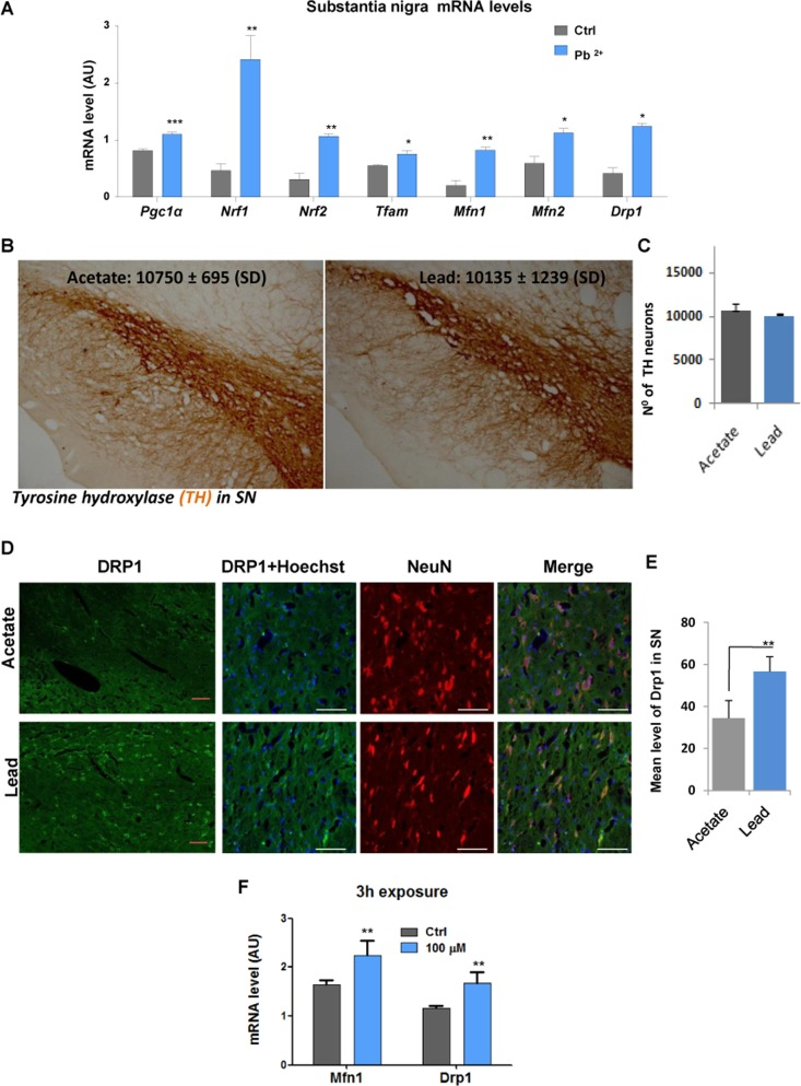
Figure 4.In vivo sub-lethallead concentration in drinking water increases the expression of genes controlling mitochondrial biogenesis and dynamicsAdult rats received drinking water containing 500 ppm lead acetate for 14 weeks and control animals received water with acetate only. Substantia nigra tissue was isolated and mRNA was extracted and processed for Real time QPCR analysis. (A) mRNA levels of Pgc1α and its target genes Nrf1, Nrf2 and Tfam as well asMfn1, Mfn2and Drp1 were analysed by qRT-PCR; (B) Immunohistochemistry was performed to detect tyrosine hydroxylase (TH) positive neurons and (C) stereological counting of TH-positive neurons in the substantia nigra of rats given drinking water containing 500 ppm lead acetate for 14 weeks. (D) Immunohistochemistry of DRP1 and NeuN co-localisation in the ventral mesencephalon neuronal cells. (E) Drp1 signal intensity was analysed by confocal laser scanning microscope (Zeiss LSM 7 DUO), using the associated software package (ZEN 2010) and quantified using an Image Analysis Program from Soft Imaging System (analySIS®, Germany). (F) MfnI and Drp1 mRNA levels in N27 cells after 3-hours of lead exposure. *P<0.05, **P<0.01, ***P<0.001, n=6 (F, n=3); mean ± SE.