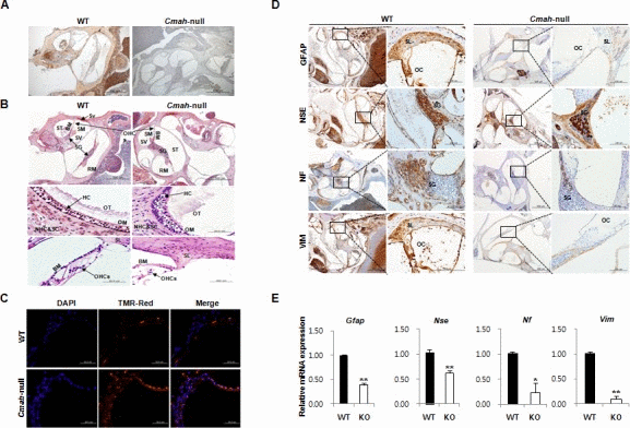
Figure 1.Age-related inner ear abnormality and neuron cell loss in Cmah -null mice(A) Analysis of Neu5Gc expression in cochlear tissues of WT- and Cmah-null mice by IHC using a chicken anti-Neu5Gc antibody. (B) Histological abnormality in inner ears of Cmah-null mice. Deposition of unusual and apparently a cellular material in the vestibular otoconial epithelia (upper). The area of the outer hair cells showed degeneration of the sensory cells throughout the cochlea in the cochlear sensory epithelium, (bottom). SV: scala vestibule, SM: scala media, ST: scala tympani, SG: spiral ganglion, BM: sasilar membrane, RM: reissner's membrane, Sv: stria vascularis, OT: otoconia, OM: otolithic membrane, NHC & SC: neuroepithelial hair cells and supporting cells, HC: hair cells, OHCs: outer hair cells, SL: Spiral lamina. (C) Apoptotic cell death in the cochlea of Cmah-null mice. TUNEL labeling performed on paraffin sections the cochlea from WT- and Cmah-null mice. (D) The expression of ganglion cells in cochlear tissues from WT- and Cmah-null mice by IHC. Rectangular box indicate a higher magnification images (5x) in left panel. GFAP: Glial fibrillary acidic protein, NSE: Neuron specific enolase, NF: Neurofilament, VIM: Vimentin. SL: spiral lamina, OC: organ of corti, SG: spiral ganglion. (E) mRNA expression pattern of neuronal cell markers in cochlear tissues from WT- and Cmah-null mice. RT-qPCR was used to measure the expression of Gfap, Nse, Nf, and Vim in the cochlea from WT- and Cmah-null mice.