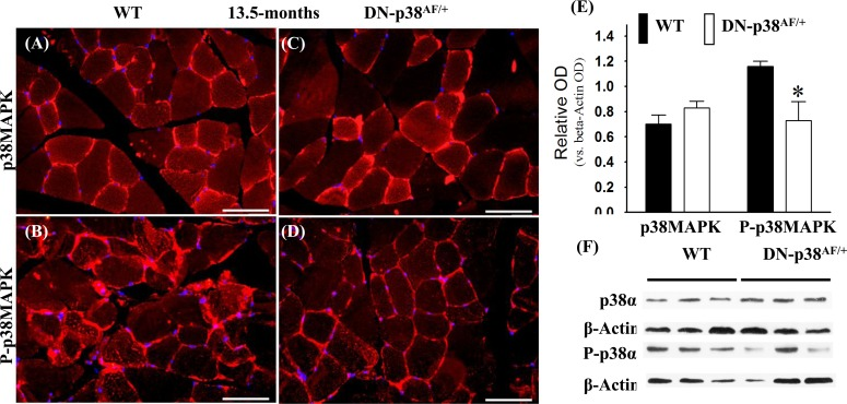
Figure 2.Expression of p38α and phospho-p38α in the gastrocnemius of middle-aged (13.5 mos old) wild type and DN-p38αAF/+ mice(A-D) Immunohistofluorescence analysis of the levels of p38α and P-p38α in cross sections of the gastrocnemius of middle aged (13.5 mos) (A, B) WT and (C, D) DN-p38αAF/+ mice. The red immunohistofluorescence depicts levels of p38α or P-p38α using antibodies specific to either p38α or P-p38α. The blue immunofluorescence depicts DAPI stained nuclei; Scale bar = 50 μm. (E) A bar graph presentation of the western blot data in (F). The data in (E) are depicted as relative OD vs. β-Actin values of Western blots. *p < 0.05, vs. corresponding WT. (F) Western blot (immunoblot) analysis of the levels of p38α pool and total P-p38α pool in (A, B) middle aged WT (13.5 mos) and (C, D) DN-p38AF/+ mice.