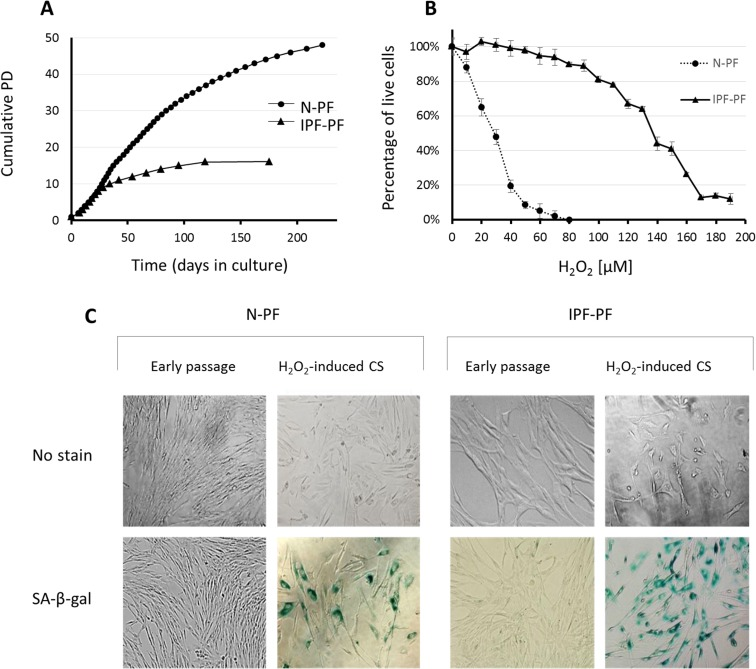
Figure 1.Growth curves and response to oxidative stress of IPF-derived and normal human pulmonary fibroblasts(A) Lung fibroblasts derived from IPF patients (IPF-PF) and healthy subjects (N-PF) during routine culture. PD stands for Population Doublings. Note that IPF-derived fibroblasts cease to proliferate after passage 16, whereas the N-PF ones are still in the logarithmic phase of cell growth. The difference between N-PF and IPF-PF growth curves is highly significant (Mann–Whitney U-test; p < 0.001). (B) Lung fibroblasts derived from IPF patients (IPF-PF) and healthy (N-PF) were treated with indicated doses of H2O2 for two hours, and tested for viability by Neutral Red assay (LD50 was 28.7 μM and 136 μM for N-PF and IPF-PF, respectively; p < E-06). Results represent 3 independent experiments. (C) SA-β-gal staining of normal (left panel) and IPF-derived fibroblasts (right panel) treated with H2O2 (the doses that caused 20% cytotoxicity were used).