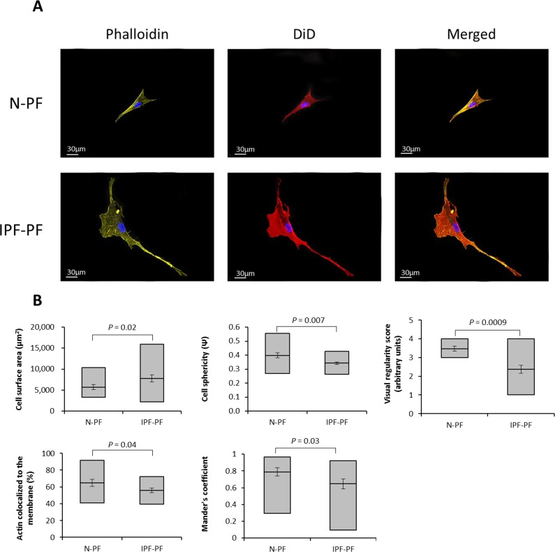
Figure 3.Morphology analysis of lung fibroblasts by Z-stack confocal microscopy(A) Representative image of lung fibroblasts derived from IPF patients (IPF-PF) and healthy (N-PF). Background subtracted. Phalloidin stains for actin; DiD for membrane and all pictures show DAPI staining of the nucleus (B) Quantification of cell morphology using the Imaris software (see methods) of confocal z-stack images taken for fibroblasts of early passage (passage 7). Whiskers indicate standard error [; bars indicate range (min/max); middle line indicates mean.