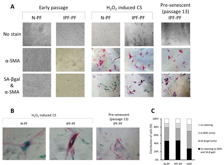
Figure 4.Immunostaining for SA-β-gal and α-SMA in primary cultures of pulmonary fibroblasts(A) early passages (upper left panels) and at cellular senescence (upper right panels). SA-β-gal – green staining; α-SMA – red staining. (B) Representative co-stained cells. (C) Distribution of cells expressing α-SMA, SA-β-gal, or both in senescent primary cultures of normal pulmonary (N-PF), IPF-derived fibroblasts (IPF-PF) and normal dermal Fibroblasts (HDF). The difference between the two pulmonary cell types was insignificant (p > 0.05).