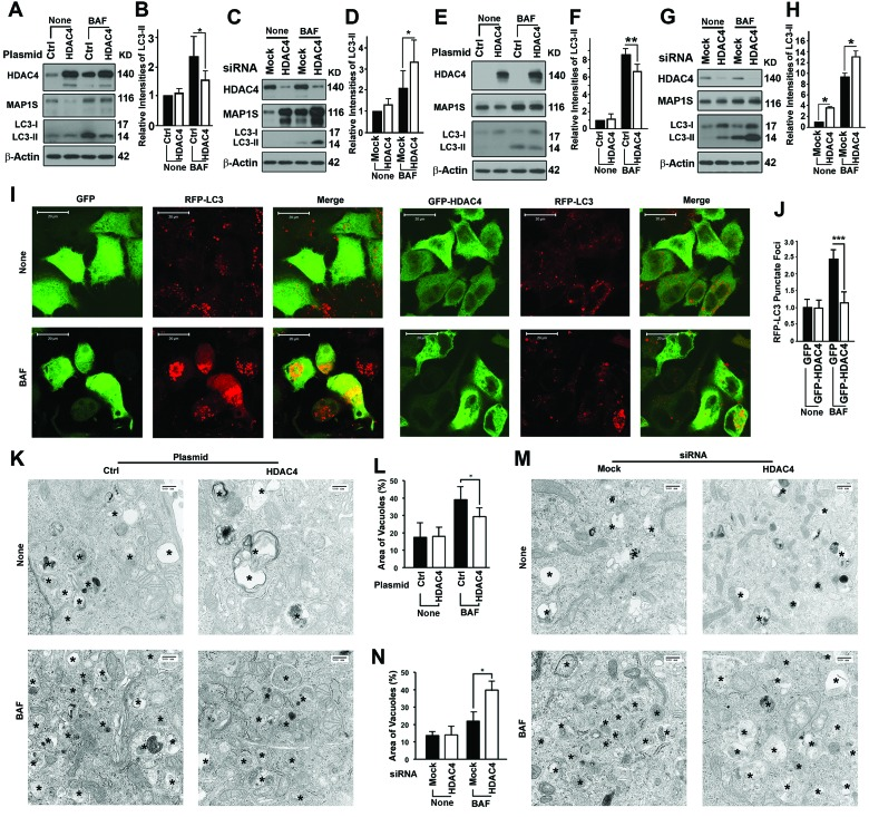
Figure 2.HDAC4 inhibits autophagy flux(A-D) HDAC4 affects levels of MAP1S and LC3-II in HeLa cells in the absence (None) or presence of BAF. Representative immunoblot results (A, C) and their respective quantification (B, D) of the impact of HDAC4 overexpression (A, B) and HDAC4 suppression with siRNA (C, D) are shown. (E-H) HDAC4 affects levels of MAP1S and LC3-II in N2a cells in the absence (None) or presence of BAF. Representative immunoblot results (E, G) and their respective quantification (F, H) of the impact of HDAC4 overexpression (E, F) or HDAC4 suppression with siRNA (G, H) are shown. (I, J) HDAC4 overexpression reduces punctate foci of RFP-LC3 in HeLa cells stably expressing RFP-LC3. Fluorescence microscopy images (I) and quantification (J) of punctate foci of RFP-LC3 in the absence or presence of BAF are shown. (K-N) HDAC4 overexpression (K, L) or siRNA suppression (M, N) alters the vacuolar areas in HeLa cells. Transmission electron microscopic images (K, M) and quantification (L, N) are shown. Symbol “*” indicates vacuoles.