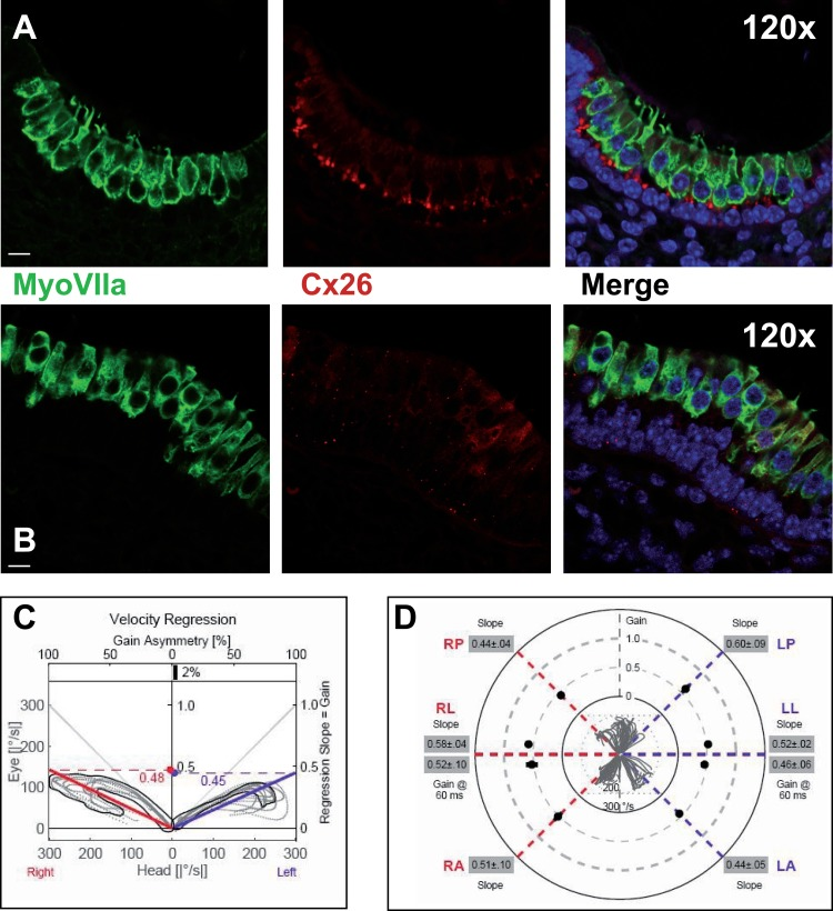
Figure 3.Macular structure in p63 defective mice and vestibular function in EEC patient(A) Representative picture of a macula of the vestibule from mice normal controls stained with MyoVIIa and Connexin 26 antibody. Dapi has been used for nuclei staining. Bars =20 μm. (B) Macular neuroepithelium from p63−/− mice. (C) Results of vHIT testing of the lateral semicircular canal (LSC) for the patient described in the article; the regression analysis of the vestibulo-ocular reflex (VOR) gain (eye angular velocity/head angular velocity) shows values of 0.48 and 0.45 for the right and left LSC, respectively. This shows VOR hyporeflectivity (average gain between 0.8 and 1.1). (D) The vestibular hyporeflectivity for both sides was confirmed by studying VOR gain for all the semicircular canals, with instant gain at 60 milliseconds for the lateral semicircular canals: gain values were all under average, without any significant asymmetry involving one side with respect to the other. Canals positioned on the same plane are connected by lines; RA= right anterior, RP= right posterior, RL=right lateral; LA= left anterior, LP= left posterior, LL=left lateral.