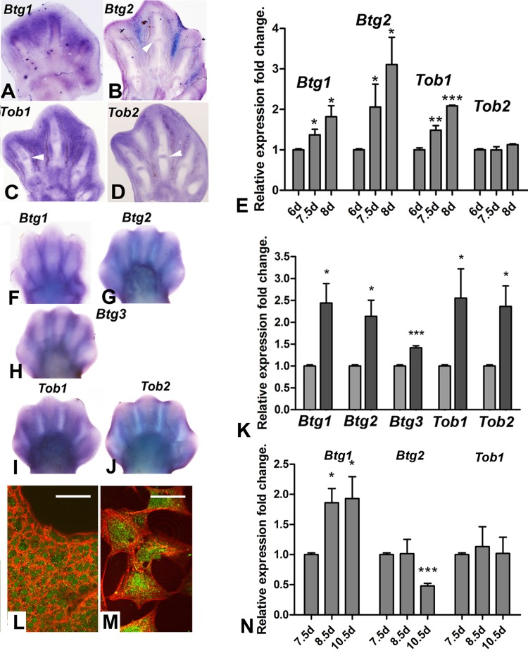
Figure 2.(A-D) in situ hybridizations showing the expression of Btg1 (A), Btg2 (B), Tob1(C) and Tob2 (D) in the chick autopod during interdigit regression. Note that, in addition to the interdigital domains, Tob2, Btg2 and Tob1 are also expressed in the developing interphalangeal joints (arrows). (E) Expression level of Btg and Tob genes in interdigital tissue of chick leg during the course of remodeling. The chart shows QPCR-evaluated fold changes in the expression of Btg1, Btg2, Tob1 and Tob2 in the third interdigit of the chick leg bud at 7.5 and 8 id compared with their expression levels prior to the onset of tissue regression (6 id). (F-J) in situ hybridizations showing the expression of the Btg/Tob genes in the developing mouse autopod. (K) chart is a comparative QPCR analysis of the interdigital expression of Btg/Tob genes at day 13 p.c. (light columns) versus day 13,5 pc (dark columns). (L-M) immunostaining for BTG2 (green) combined with phalloidin (red) in vibratome sections of the third interdigit (M), and in cultured mesodermal progenitors (M). Note that the protein is expressed in the cytoplasm and nuclei. Scale bar in L = 100μm; Bar in M = 20μm. (N) shows a QPCR analysis of the expression of Btg1, Btg2 and Tob1 in the third interdigit of embryonic duck leg at equivalent stages of that of the chick in E. Unlike in the chick (compare with E), Btg2 becomes down-regulated and Tob1 is not up-regulated over the course of tissue remodeling. ***p < 0,001; ** p < 0.01; * p< 0.05.