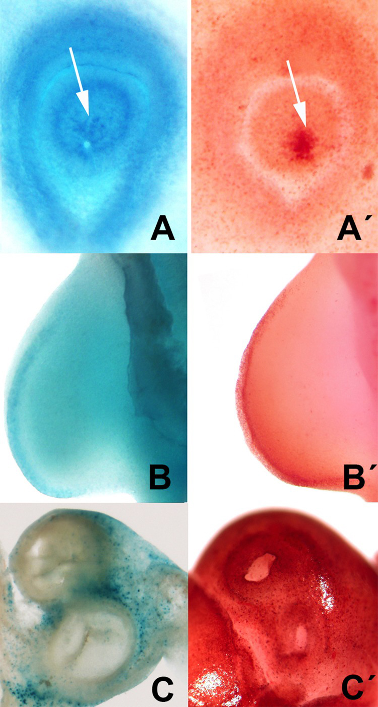
Figure 6.Selection of areas of embryonic programmed cell death showing the parallelism between the distribution of neutral red vital staining (A′, B′, C′) and β-gal activity (A, B C). A-A′: cell death (arrows) during the closure of the lens in chick embryos at 2.5 id. B-B′ cell death in the AER in the embryonic limb at id 3.5. C-C′, cell death in the root of the main arteries of the embryonic heart at id 7.5.