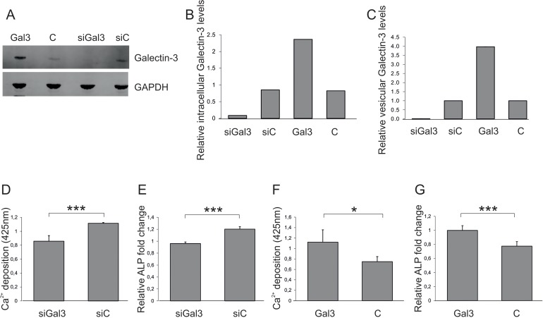
Figure 3.Impact of vesicular Galectin-3 levels on osteogenic commitment of ASCs(A) Detection and (B) quantification of total Galectin-3 protein levels normalized to GAPDH by Western blot in protein lysates derived from endothelial cells (ECs) transfected with siRNA against Galectin-3 (siGal3), a Galectin-3 overexpression construct (Gal3) or the corresponding controls (siC or C). (C) Galectin-3 levels of extracellular vesicles isolated from endothelial cells (ECs) transfected with siRNA against Galectin-3 (siGal3), a Galectin-3 overexpression construct (Gal3) or the corresponding controls (siC or C) were analysed by ELISA and normalized to the number of donor cells (D-E) ASCs were exposed to EVs isolated from siRNA against Galectin-3 (siGal3) or corresponding non-targeting control (siC) transfected endothelial cells. Osteogenic differentiation was reduced in ASCs co-incubated with vesicles derived from HUVECs expressing less Galectin-3 (siGal3) as compared to cells exposed to extracellular vesicles of control transfected HUVECs (siC) as evaluated by (D) Alizarin Red staining and (E) qPCR on ALP mRNA levels normalized to GAPDH. (F-G) ASCs exposed to EVs of Galectin-3 expression plasmid (Gal3) or empty vector (C) transfected HUVECS. Osteogenic differentiation was enhanced in ASCs co-incubated with vesicles derived from Galectin-3 overexpressing HUVECs as compared to cells exposed to extracellular vesicles of empty vector transfected HUVECs as quantified by (F) was Alizarin Red staining and (G) qPCR on ALP mRNA levels normalized to GAPDH. (D-G) *: p<0.05, ***: p<0.001 in comparison to control. Data are presented as mean values ± SD and were statistically analysed using unpaired t test, n=4.