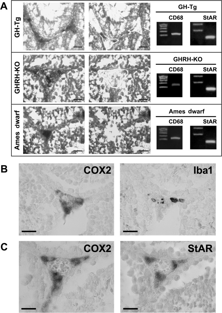
Figure 2.COX2 is expressed in testicular macrophages and Leydig cellsPanel (A) COX2-immunoreactive interstitial cells were isolated by Laser Capture Microdissection (LCM) from testicular sections of short-lived (GH-Tg) and long-lived (Ames dwarf and GHRH-KO) mice. The same section is illustrated before (left panel) and after (right panel) LCM. Bar, 25 μm. A total of 50 to 80 COX2-immunopositive interstitial cells were isolated by LCM and subsequently used to evaluate the expression of CD68 (macrophage cell marker) and StAR (Leydig cell marker) by RT-PCR. Panels (B and C) Immuno-colocalization of COX2 and Iba1 (macrophage cell marker; Panel (B) and immuno-colocalization of COX2 and StAR (Leydig cell marker; Panel (C) in testicular sections from a short-lived mouse (GH-Tg) was examined using a light microscope. Bar, 20 (m. Similar images were seen when testicular sections from long-lived (Ames dwarf and GHRH-KO) mice were used (data not shown).