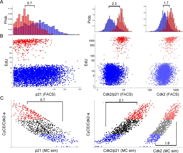
Figure 8.Single cell proliferation analysis of G1-S (2n) cells 3 days after 2.5 Gy IR. Cells were stained for DNA content, p21, Cdk2 and EdU. Only single 2n gated cells are shown. X-scales of A and B are equal. (A) Probability distribution histogram for EdU-positive (red) and EdU-negative (blue) cells. Corresponding dot plots are displayed in B. Numbers indicate the fold-difference of population means. Left panel: EdU vs. p21, Right panel: EdU vs. Cdk2/p21 and Cdk2, respectively, as indicated on the x-axis in B. (B) Dot plots of single cell EdU incorporation. Red: EdU-positive cells, Blue: EdU-negative cells. Left panel: EdU vs. p21, Right panel: EdU vs. Cdk2/p21 (bright dots) and Cdk2 (shaded dots), respectively. Cdk2/p21 and Cdk2 are displayed in the same graph to illustrate distribution differences. (C) Single cell Monte-Carlo simulation (MC sim) 3 days after 2.5 Gy IR. Red: Upper 25%-quantile of simulated active Cdk2 (CycE/Cdk2-a), Blue: Lower 25%-quantile of simulated active Cdk2 (CycE/Cdk2-a). Left panel: CycE/Cdk2-a vs. p21, Right panel: CycE/Cdk2-a vs. Cdk2/p21 (bright dots) and Cdk2 (lighter dots), respectively. Cdk2/p21 and Cdk2 are displayed in the same graph to illustrate distribution differences. Numbers indicate the fold-difference between red and blue population means.