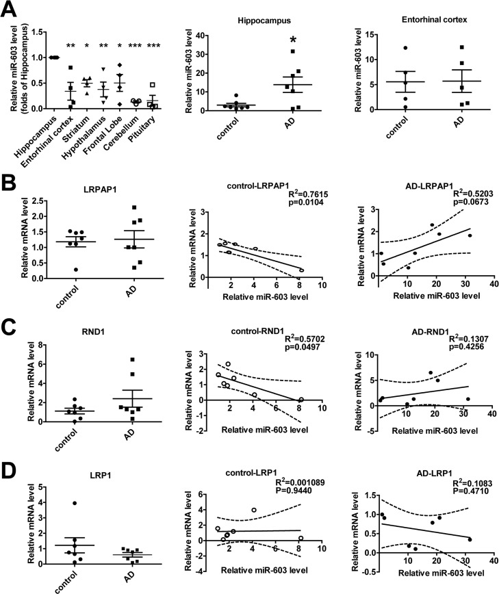
Figure 6.miR-603 exhibits a relatively higher expression in the hippocampi of patients with AD(A) Quantitative RT-PCR analysis of miR-603 expression in brain samples (n = 4 for each group, left), *P < 0.05, **P < 0.01, ***P < 0.001 versus the hippocampus as determined by one-way ANOVA with a Dunnett multiple comparison test. Quantitative RT-PCR analysis of miR-603 expression in the hippocampi (middle) and entorhinal cortexes (right) of control subjects and patients with AD (n = 7 for each group); *P < 0.05 as determined by an unpaired two-tailed Student's t-test. (B) Quantitative RT-PCR analysis of LRPAP1 mRNA levels in the hippocampi of control subjects and patients with AD (n = 7 for each group, left) and linear regression analysis of the correlation between miR-603 expression and LRPAP1 mRNA levels in control groups (middle) and AD groups (right). (C) Quantitative RT-PCR analysis of RND1 mRNA levels in the hippocampi of control subjects and patients with AD (n = 7 for each group, left) and linear regression analysis of the correlation between miR-603 expression and RND1 mRNA levels in control groups (middle) and AD groups (right). (D) Quantitative RT-PCR analysis of LRP1 mRNA levels in the hippocampi of control subjects and patients with AD (n = 7 for each group, left) and linear regression analysis of the association between miR-603 expression and LRP1 mRNA levels in control groups (middle) and AD groups (right). All the subjects were identified by allocated numbers. The miR-603 expression and LRPAP1, RND1, LRP1 mRNA levels of each subject were standardized by the corresponding values of subject No. 78.