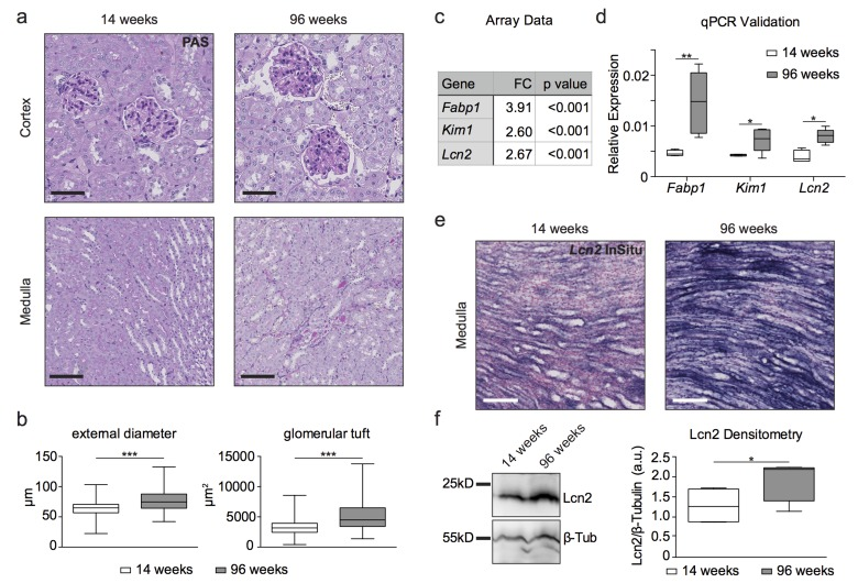
Figure 1.Histology, transcriptome analysis, western blot and in situ hybridization reveal a kidney aging phenotype(a) PAS staining of young and aged wildtype kidneys. Aged kidneys show cysts and hypertrophic glomeruli, prominent basal membranes and dilated capillary loops in the renal cortex as well as protein cylinders in parts of the medulla. Scale bars - upper panel: 50μm; lower panel: 100μm (b) Quantitative measurement of glomeruli by their external diameter and glomerular tuft area. Aged glomeruli show a hypertrophy compared to 14 week old glomeruli. (c) Table of fold change (FC) in kidney damage markers obtained from microarray analysis. (d) qPCR validation of array data for kidney damage markers. (e) In situ hybridization for Lcn2 (NGAL)-RNA on formalin-fixed paraffin-embedded kidney tissue. 96 week old kidneys show increased Lcn2 RNA levels in the papilla compared to young animals. Scale bar: 100μm (f) Immunoblot for Lcn2 shows a clear increase in protein content in 96 week old kidney lysates. β-tubulin was used as a loading control and for normalization of densitometry. Boxplots depict mean values with whiskers showing 5-95% percentile.*p<0.05, **p<0.01, ***p<0,001.