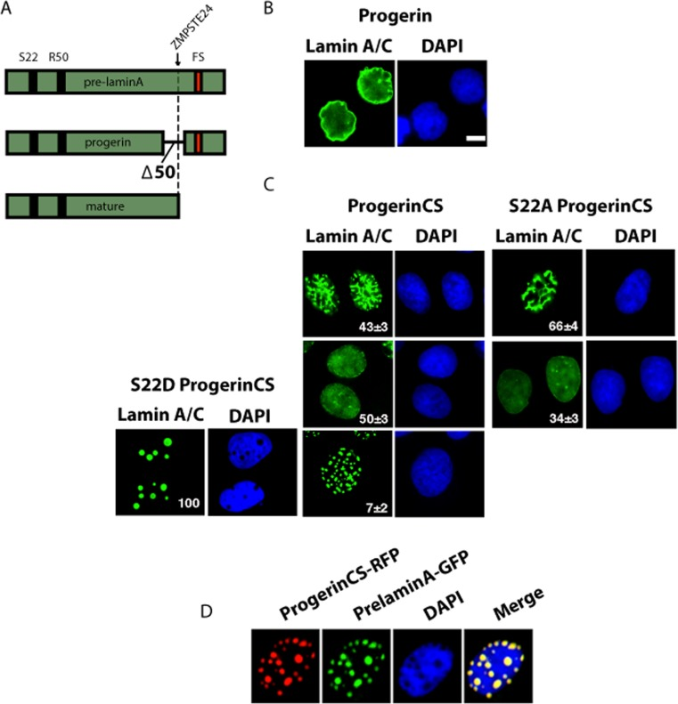
Figure 1.Distinct patterns of nuclear localization of progerin and non-farnesylated progerin (progerin CS)(A) Schematic representation of prelamin A alleles cloned into retroviral vectors and used in this study. FS= farnesylation site. Note the ZMPSTE24 cleavage site (arrow) that is absent in progerin. (B-C) Immunofluorescence for lamin A/C of U-2 OS cell expressing progerin or progerin CS with wild type S22 or mutations S22A or S22D. Cells were fixed 5 days after infection. The percent and standard deviation (S.D) of cells having each pattern is indicated at the bottom right of each panel. (D) Immunofluorescence images of U-2 OS cell coexpressing progerin CS fused with Red fluorescent protein (RFP) and prelamin A fused with GFP. Magnification = 10 μm.