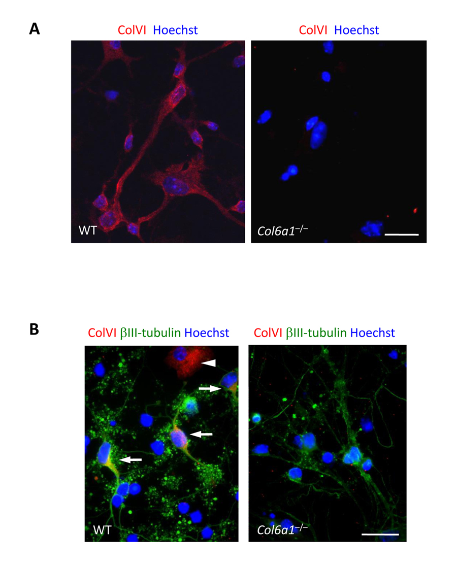
Figure 1.Collagen VI immunolabeling in primary neural cell cultures(A) Confocal microscopy analysis of immune-fluorescence for collagen VI in primary neural cell cultures. Collagen VI (red) is detected in wild-type, but not in Col6a1−/− cultures. Nuclei were stained with Hoechst (blue). Scale bar, 100 μm. (B) Immunofluorescence for collagen VI (red) and βIII-tubulin (green). In wild-type cultures, collagen VI labelling is detected in both neuronal (βIII-tubulin-positive, arrows) and glial (βIII-tubulin-negative, arrowhead) cells. Nuclei were stained with Hoechst (blue). Scale bar, 100 μm. ColVI, collagen VI; WT, wild-type.