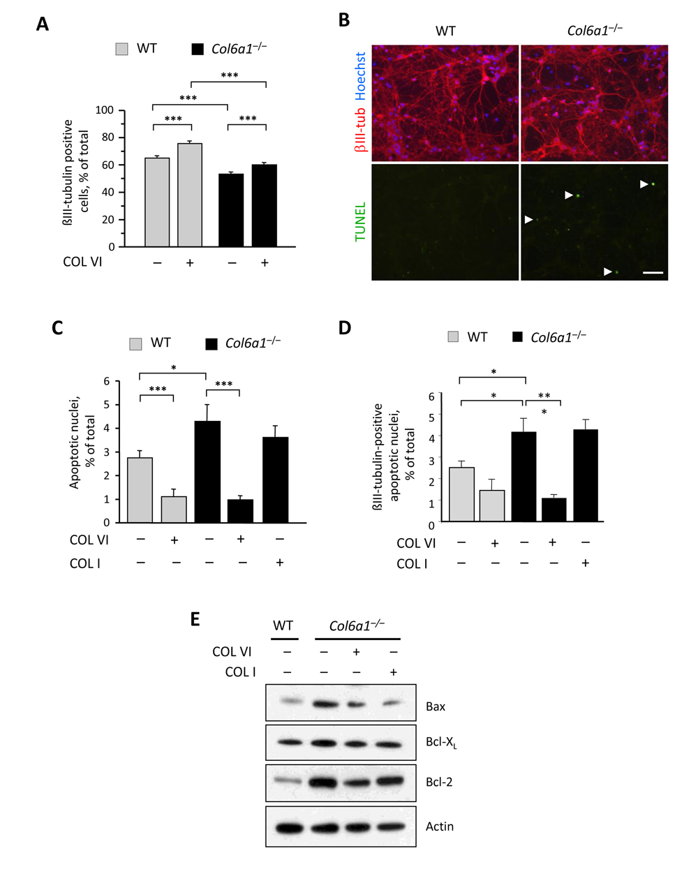
Figure 2.Lack of collagen VI affects the neuronal population and induces increased apoptosis in neural cell cultures(A) Quantification of the percentage of βIII-tubulin-positive cells in primary neural cell cultures derived from wild-type and Col6a1−/− mice, grown in the absence (−) or in the presence (+) of purified collagen VI as a substrate (***, P<0.001; n = 6). (B) Representative images of TUNEL analysis in primary neural cell cultures derived from wild-type and Col6a1−/− mice. Neurons were stained by immunofluorescence for βIII-tubulin (red, upper panels), and nuclei were labelled with Hoechst (blue, upper panels). TUNEL-positive nuclei are green (arrowheads, lower panels). Scale bar, 50 μm. (C,D) Quantification of TUNEL-positive nuclei in all cells (C) and of TUNEL-positive nuclei in βIII-tubulin-positive cells (D) in primary neural cell cultures derived from wild-type and Col6a1−/− mice, grown in the absence (−) or in the presence (+) of collagen VI or collagen I as substrates (***, P<0.001; *, P<0.05; n = 3). (E) Western blot analysis of pro- and anti-apoptotic factors in total protein extracts derived from wild-type and Col6a1−/− primary neural cell cultures. Actin was used as a loading control. COL I, purified collagen I; COL VI, purified collagen VI; WT, wild-type.