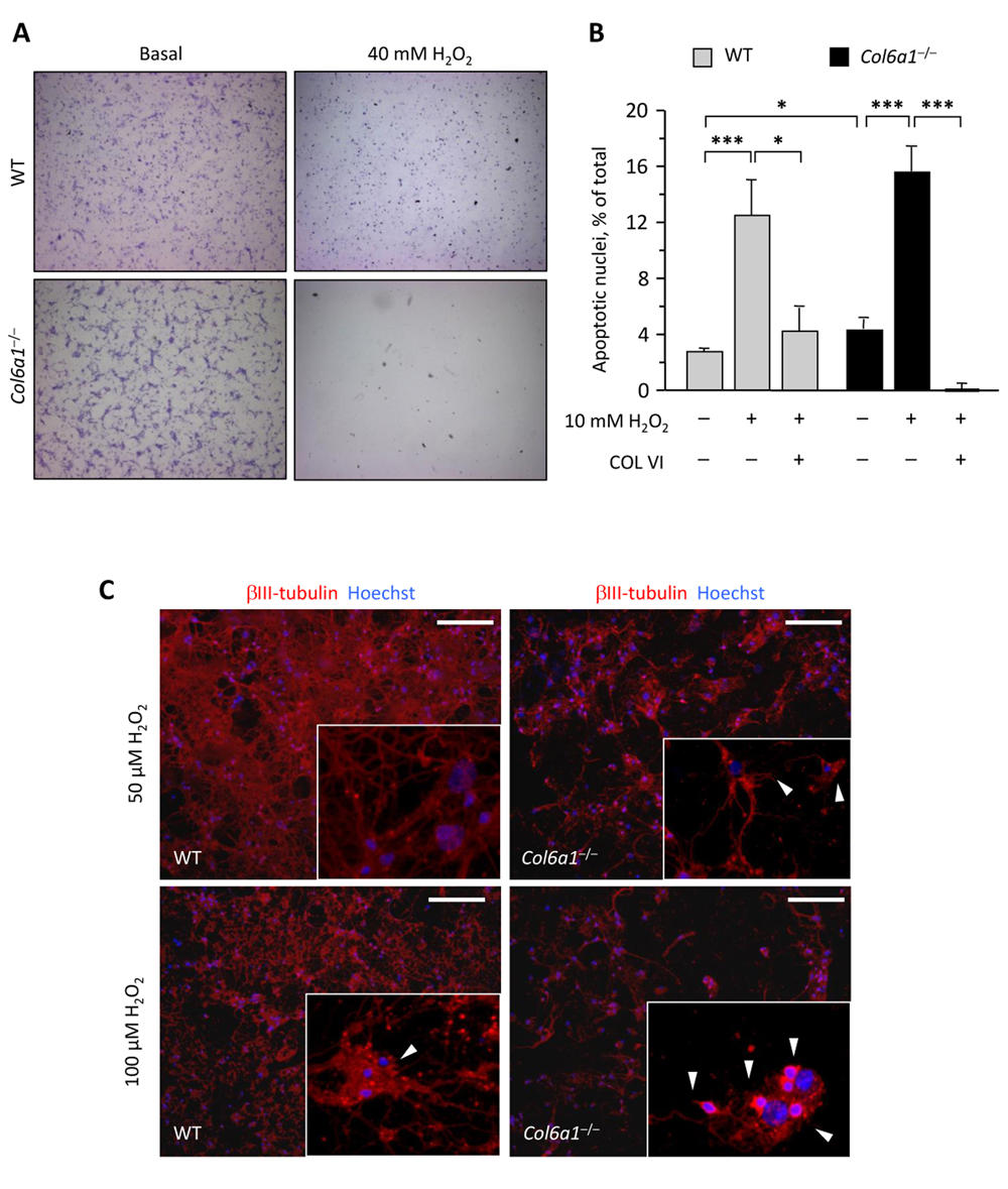
Figure 4.Col6a1−/− neural cell cultures display higher oxidative damage(A) Light microscopy analysis of wild-type and Col6a1−/− primary neural cell cultures maintained in standard condition (Basal) or after treatment for 90 min with 40 mM H2O2. Cells were fixed and stained with cresyl violet. Scale bar, 100 μm. (B) Quantification of TUNEL-positive nuclei in wild-type and Col6a1−/− primary neural cell cultures maintained in standard condition or after treatment for 90 min with 10 mM H2O2. Where indicated, cells were grown onto purified collagen VI before treatment (***, P < 0.001; *, P<0.05; n = 3). COL VI, adhesion onto purified collagen VI. WT, wild-type. (C) Immunofluorescence for βIII-tubulin (red) in wild-type and Col6a1−/− neural cell cultures after treatment for 90 min with 50 μM H2O2 (top panels) or 100 μM H2O2 (bottom panels). Even at lower doses of hydrogen peroxide, Col6a1−/− cultures display a less dense neuronal network, with higher incidence of dendrite shrinkage (arrowheads in insets). The insets show higher magnification details of each panel. Nuclei were stained with Hoechst (blue). Scale bar, 50 μm. WT, wild-type.