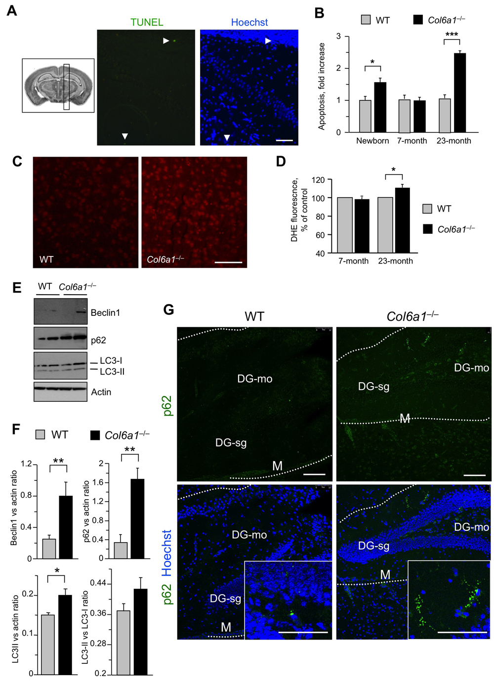
Figure 6.Aged Col6a1−/− mice display neurodegenerative hallmarks(A) Representative image of TUNEL assay in adult Col6a1−/− mouse brain sections derived from the region showed in the inset on the left. TUNEL-positive nuclei (green) are shown in the middle panel. Nuclei were stained with Hoechst (blue, right panel). Scale bar, 50 μm. (B) Quantification of TUNEL-positive nuclei in sagittal sections of brains of newborn, 7-month-old and 23-month-old wild-type and Col6a1−/− mice. The number of TUNEL-positive cells was counted per area unit (mm2), and the incidence of apoptosis in Col6a1−/− mice calculated as fold-change relative to the age-matched wild-type values (***, P<0.01; *, P<0.05; n = 3-7). (C) Representative images of DHE staining in brain sections of 23-month-old wild- type and Col6a1−/− mice. Scale bar, 100 μm. (D) Quantification of DHE fluorescence in brain sections of 7-month-old and 23-month-old wild-type and Col6a1−/− mice. Values for Col6a1−/− mice are reported as percentage relative to the age-matched wild-type value. ROS accumulation is significantly higher in aged Col6a1−/− mice (*, P<0.05; n = 3-7). (E) Western blot analysis of the autophagic markers Beclin 1, p62 and LC3 in total protein extracts from brain of 23-month-old wild-type and Col6a1−/− mice. Actin was used as a loading control. (F) Densitometric quantification of Beclin 1/actin ratio, p62/actin ratio, LC3-I/actin ratio and LC3-II/LC3-I ratio, as determined by three independent western blot experiments of brain extracts from 23-month-old wild-type and Col6a1−/− mice. (**, P < 0.01; *, P<0.05; n = 3). (G) Immunofluorescence for p62 in brain sections from 23-month-old wild-type and Col6a1−/− mice, revealing increased p62 labeling (green) in Col6a1−/− samples. Insets show p62 aggregates at higher magnifications. Scale bar, 100 μm. DG-mo, dentate gyrus, molecular layer; DG-sg, dentate gyrus, granule cell layer; M, meningis; WT, wild-type.