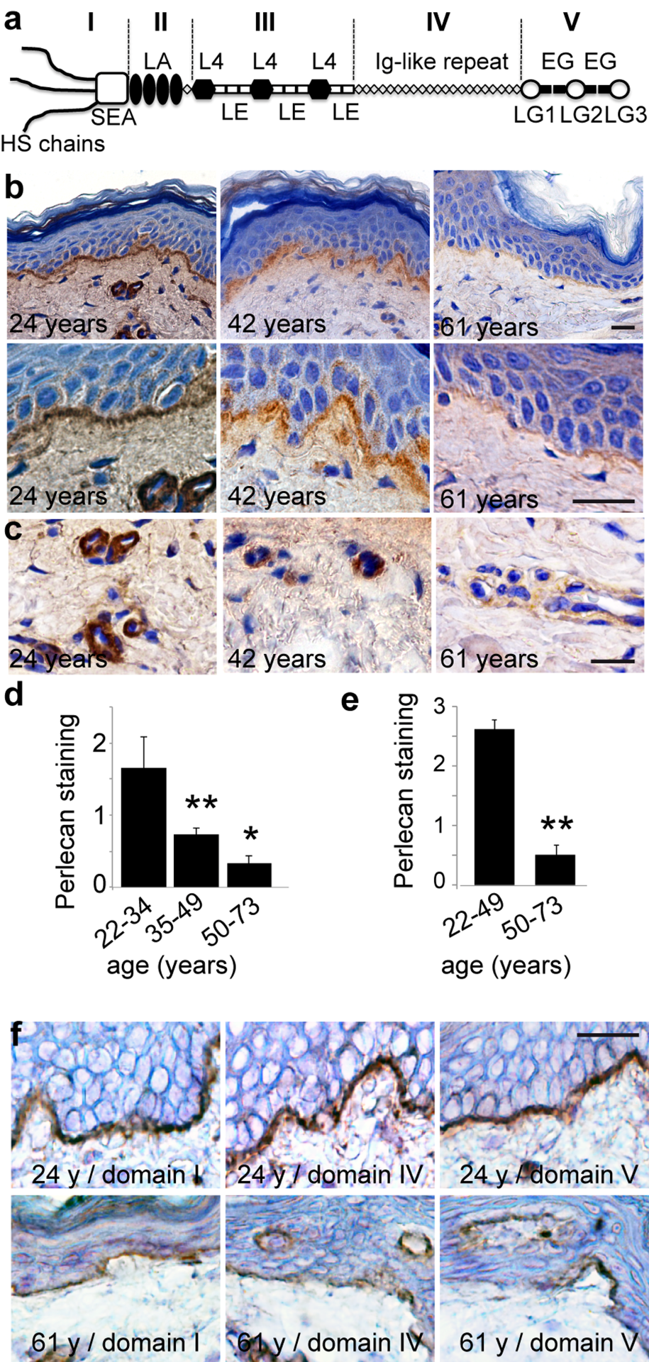
Figure 1.Age‐related changes in perlecan expression in skin basement membranes(a) Schematic representation of perlecan. (b, c) Perlecan expression in skin from women donors aged 24, 42, and 61 years (domain III stain). Sections of paraffin‐embedded skin biopsies were stained for perlecan and counterstained with Harris haematoxylin. (d, e) Quantification of perlecan staining in (d) epidermal and (e) vascular BMs in 38 human skin samples from donors of ages ranging from 22 to 73 years (22‐33, n=8; 35‐49, n=12; 50‐73, n=18). Mean ± SD, **p<0.01 vs. young group, based on Student's t‐test. (f) Age‐related reductions in perlecan subdomains. Sections of frozen skin from donors aged 24 and 61 years were immunolabelled with antibodies specific for perlecan domain I, domain IV, or domain V, and counterstained with Harris haematoxylin. (b, c, f) Scale bar = 20 μm