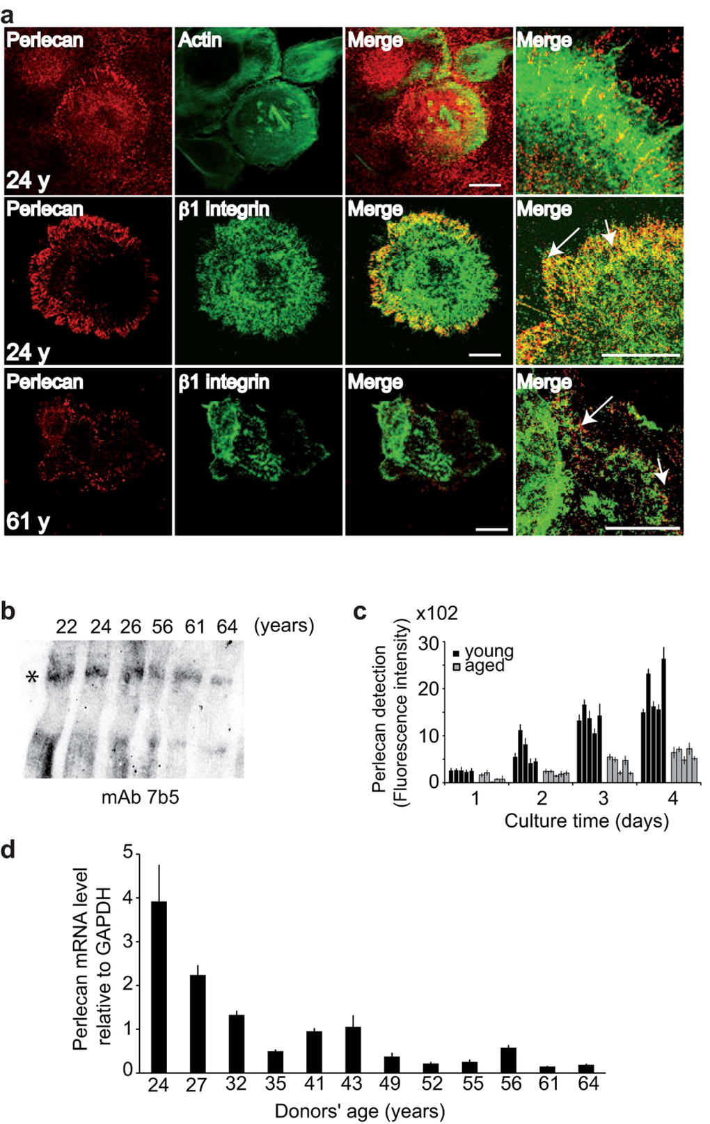
Figure 2.Keratinocyte aging results in decreased perlecan in the ECM(a) NHKs from young or aged donors were cultured, fixed, and stained with either polyclonal antibodies (pAb) against perlecan (red), phalloidin‐FITC (green) or monoclonal antibodies (mAb) against Δ1‐integrin (green), as indicated. The merged images show the juxtaposition of perlecan and β1‐integrin staining (arrows). Optical slices (0.8 μm) were acquired at the cell‐matrix interface. Scale bar = 50 μm. (b) Perlecan immunoblotting in the ECM. Ages are indicated, and the asterisk shows perlecan. (c) Perlecan quantification. NHKs (5 × 10³) from donors aged 22, 24, 26, 27, and 32 years or 52, 55, 56, 61, and 64 years were seeded in 96‐well plates, and perlecan immunodetection was performed over 4 days. Six assay points were performed per experiment. (d) Results from QPCR of NHK cDNA show perlecan gene transcription in 12 NHK strains from donors aged 24 to 64 years, as indicated. Each assay point was performed in triplicate (c, d). Data are presented as mean ± SD of 3 independent experiments conducted with keratinocytes from each individual donor.