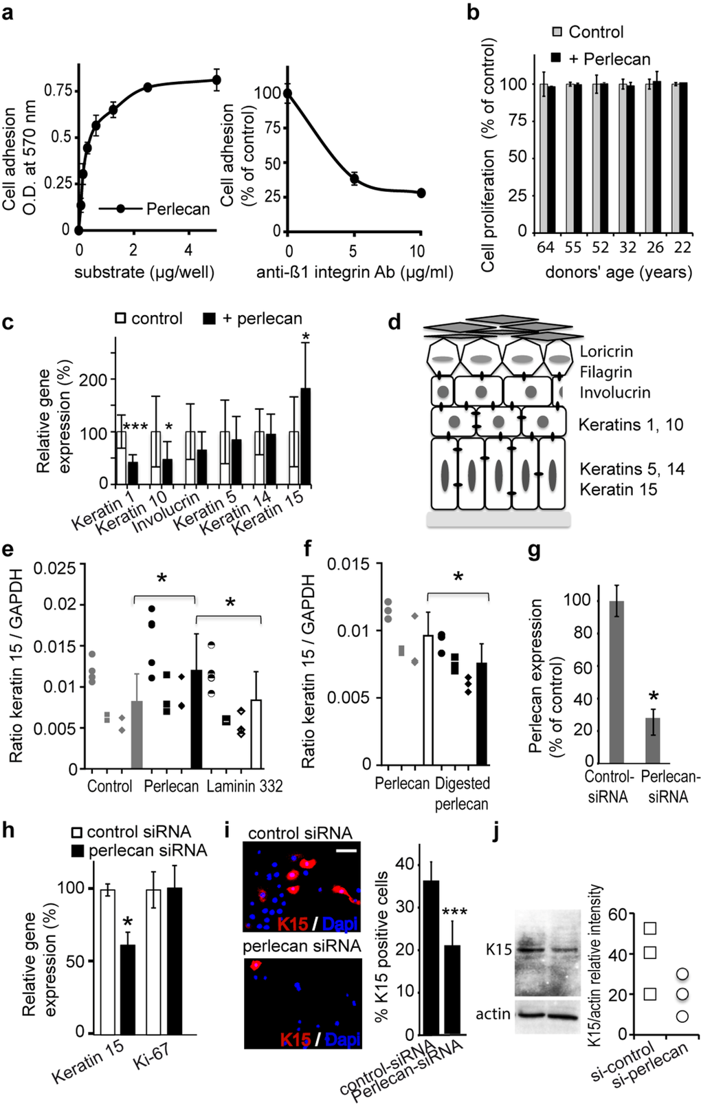
Figure 3.Perlecan induces keratinocyte adhesion and regulates K15 gene (KRT15) expression(a) Keratinocytes adhere to perlecan in a dose‐dependent manner (left). Adhesion to perlecan is inhibited by antibodies against the Δ1 integrin subunit (right). (b) Proliferation of aged and young keratinocyte strains over 24 h. The proliferation of cells plated on perlecan is expressed as a percentage of the proliferation observed in control conditions. (c) Real‐time PCR analysis of keratins (K1, K5, K10, K14, and K15) and involucrin gene expression normalized to GAPDH in keratinocytes from the 61‐year‐old donor plated on culture dishes either covered beforehand with perlecan or untreated. (d) Scheme of the epidermis showing the locations of the differentiation markers. (e) Real‐time PCR analysis of KRT15 in keratinocytes from 3 distinct aged donors (age 64, 61, 60), cultured on perlecan, laminin 332 or plastic. (f) Real‐time PCR analysis of KRT15 in keratinocytes from 3 distinct aged donors (age 64, 61, 60) cultured either on native perlecan or digested perlecan, which lacked heparin sulfate (HS) moieties. (g) ELISA results show expression levels of perlecan in the ECM of keratinocytes transfected with perlecan‐specific siRNA, compared to cells transfected with control‐siRNA. (h) Real‐time PCR analysis of KRT15 and Ki‐67 gene (MKI67) in a 52 year‐old keratinocyte strain transfected with perlecan‐specific siRNA. (i) K15 staining in perlecan‐siRNA‐transfected keratinocytes; quantification is expressed as a percentage of DAPI positive cells. Each slide was performed in triplicate; 400 cells were counted within 4 areas of each slide. (j) Western blot of K15 expression in cultured perlecan knock‐down keratinocytes. Quantification of K15 expression is relative to actin expression. Experiment was done in triplicate. (c, g, h) Data are presented as the mean ± SD of 9 independent experiments with keratinocytes from a 61 (c) or 52 (g, h) years old donor. (e, f) All performed independent experiments per donor are shown (age 64 ○, 61 □, 60 ◇) and were combined for statistical analysis. *p<0.025,**p<0.005,***p<0.0002 vs. control, Student's t‐test.