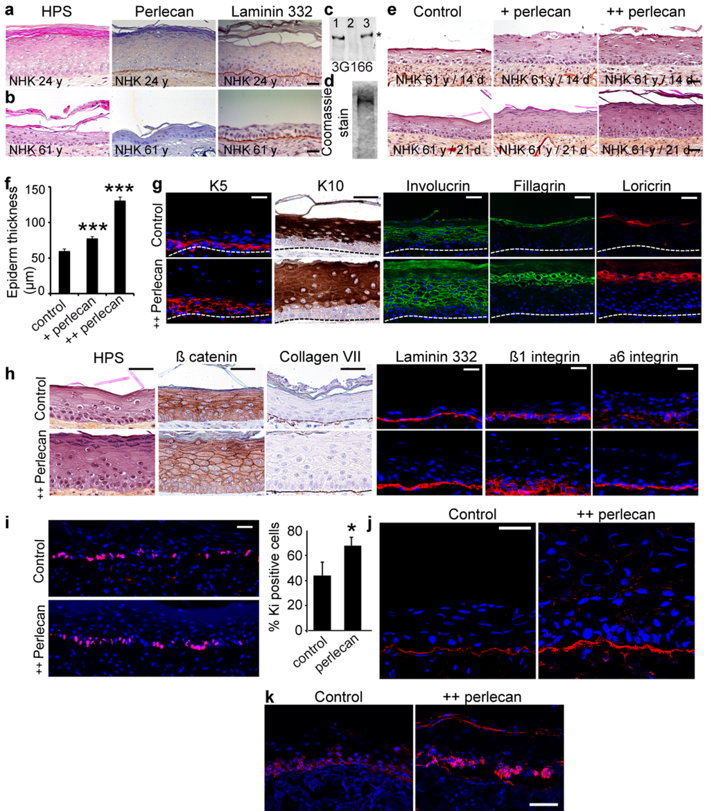
Figure 5.Exogenous soluble perlecan improves epidermal thickness(a, b) HPS staining and immunohistochemical analysis of paraffin‐embedded SEs epidermalized with (a) young or (b) aged keratinocytes. (c) Western blot of HUAEC culture medium before (lane 1) and after (lane 2) immunopurification. The eluted material (lane 3) contained the perlecan core protein. (d) Coomassie blue staining of purified perlecan. (e) SEs generated with aged keratinocytes without (control) or with perlecan (+ perlecan and ++ perlecan). SEs were collected 14 and 21 days post‐epidermalization and processed for histology. (f) Epidermal thickness. *** p<0.001 for + perlecan and ++ perlecan vs. control, Student's t‐test. (g, h) Paraffinembedded and frozen SEs at 21 days post‐epidermalization were immunolabelled to identify (g) differentiation markers and (h) adhesion/ECM components. Nuclei were stained with DAPI. (i) Ki‐67 staining in SEs. Quantification is expressed as the percentage of DAPI staining in the basal layer. Mean ± SD; *p<0.025. (j, k) Frozen SEs at 21‐days post‐epidermalization were immunostained to identify perlecan (j) and K15 (k). Scale bars = 50 μm (j), = 60 μm (k).