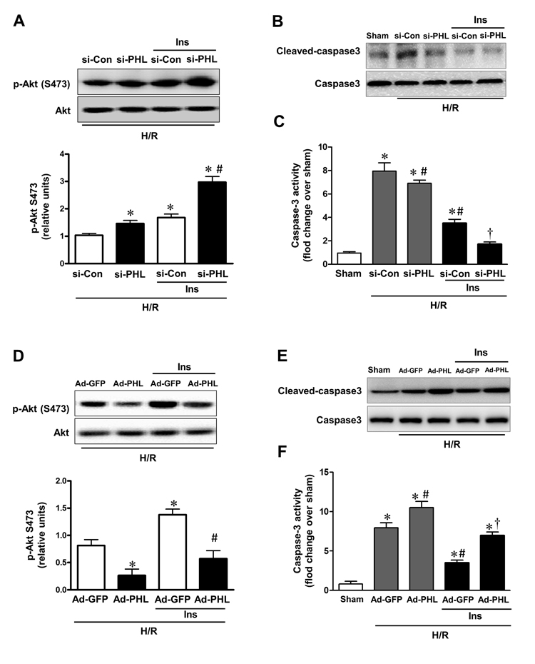
Figure 4.PHLPP-1 expression level affected insulin-activated Akt phosphorylation(A) Cardiomyocytes were transfected with PHLPP-1-specific (si-PHL) and scrambled negative control (si-Con) siRNA for 48 hours. Each group myocytes were subjected to hypoxia (3 h)/reoxygenation (3 h) with or without insulin treatment (100 nM). Representative immunoblots and bar graphs (right panel) show the relative levels of Akt phosphorylation (S473) (*P < 0.05 vs. Ad-sh-Con+H/R; #P < 0.05 vs. Ad-sh-Con+H/R+Ins). (B) Immunoblotting of cleaved/total caspase 3. (C) Quantification of caspase-3 activity in H/R-induced cardiomyocytes of the indicated group (*P < 0.05 vs. sham; #P < 0.05 vs. si-Con+H/R; †P < 0.05 vs. si-Con+H/R+Ins). (D) Cardiomyocytes were infected with Ad-PHL or Ad-GFP for 72 hours. Each group myocytes were subjected to hypoxia (3 h)/reoxygenation (3 h) with or without insulin treatment (100 nM). Immunoblots showing the relative levels of Akt phosphorylation (S473) (*P < 0.05 vs. Ad-GFP+H/R; #P < 0.05 vs. Ad-GFP+H/R+Ins). (E) Immunoblotting of cleaved/total caspase 3. (F) Quantification of caspase-3 activity in H/R-induced cardiomyocytes of the indicated group (*P < 0.05 vs. sham; #P < 0.05 vs. Ad-GFP+H/R; †P < 0.05 vs. Ad-GFP+H/R+Ins. Values are means ± S.E., n = 5 per group).