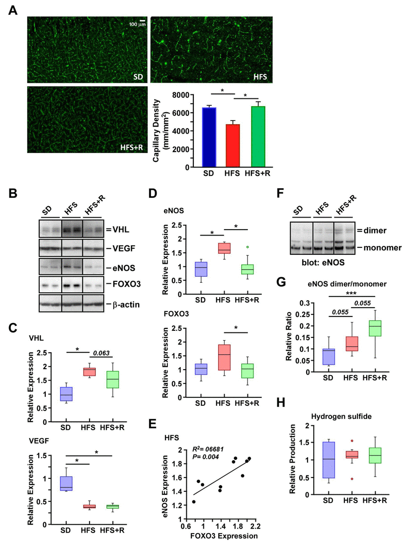
Figure 3.Resveratrol treatment improves capillary density in the cerebral cortex of HFS-fed rhesus monkeys(A) Representative images of cerebral cortical brain capillary staining, and their respective quantification. (B) Solubilized cerebral cortex extracts from SD-, HFS- and HFS+R-fed animals were resolved by SDS-PAGE under reducing conditions, electrotransferred onto nitrocellulose membranes and subjected to immunoblotting using the indicated primary antibodies. Representative signals associated with bands of interest are shown, including that of b-actin, which was used as loading control. All 24 brain samples were ran on a single gel (Fig. S1). Nitrocellulose membrane staining was also carried out with Ponceau S dye for protein detection. (C) Signals associated with VEGF and VHL proteins were normalized and represented as box plots. (D) Signals associated with eNOS and FOXO3 proteins were normalized and represented as box plots. (E) Scatterplot exploring the significant association between eNOS and FOXO3 protein expression with HFS feeding. (F) SDS-PAGE was performed at 4°C to enable the separation of eNOS dimers and monomers. Immunoblot analysis was performed using anti-eNOS antibody. (G) Ratios of dimeric and monomeric eNOS species were calculated and represented as box plots. (H) Measurement of H2S levels in monkey brain homogenates. SD (n=4); HFS (n=10); HFS+R (n=10). *, p < 0.05.