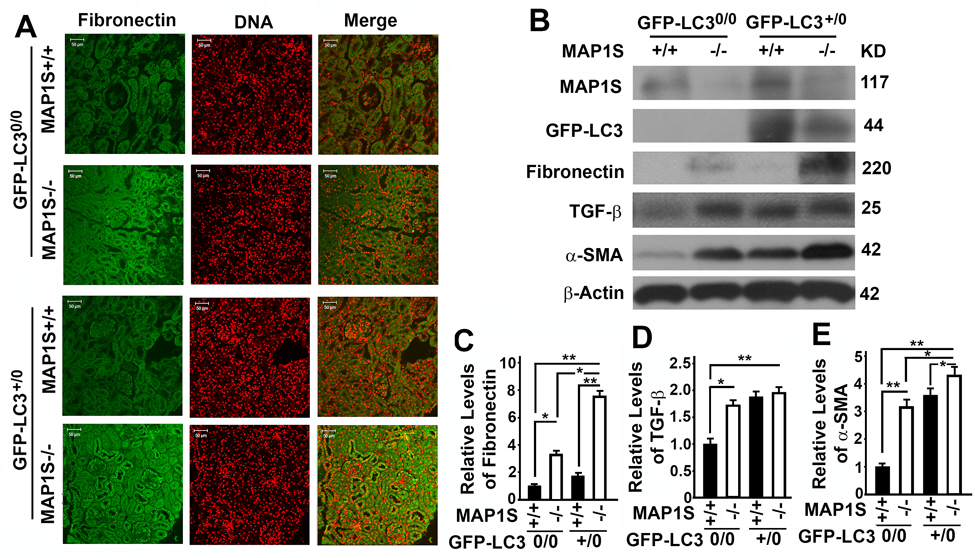
Figure 4.Autophagy defects triggered by MAP1S deficiency cause accumulation of fibronectin in mouse renal tissues(A) Immunostaining analyses of fibronectin (green) in renal tissues collected from 6-month-old wild-type (MAP1S+/+:GFP-LC30/0), knockout (MAP1S−/−:GFP-LC30/0), GFP-LC3 transgenic (MAP1S+/+:GFP-LC3+/0) and MAP1S−/−:GFP-LC3+/0 mice using anti-fibronectin antibody. Nuclear DNA is counter-stained as red. Bar: 50 μm. (B-E) Representative immunoblot images (B) and plots (C-E) showing the impacts of MAP1S on the levels of fibronectin (C), TGF-β (D) and α-SMA (E) in renal tissues described in panel (A). The initial intensity of each protein in the wild-type was set to be 1. Data shown in plots above were the averages and standard deviations of three repeats. Plots were the means ± S.D. of three repeats and the significance of the differences was compared as described above.