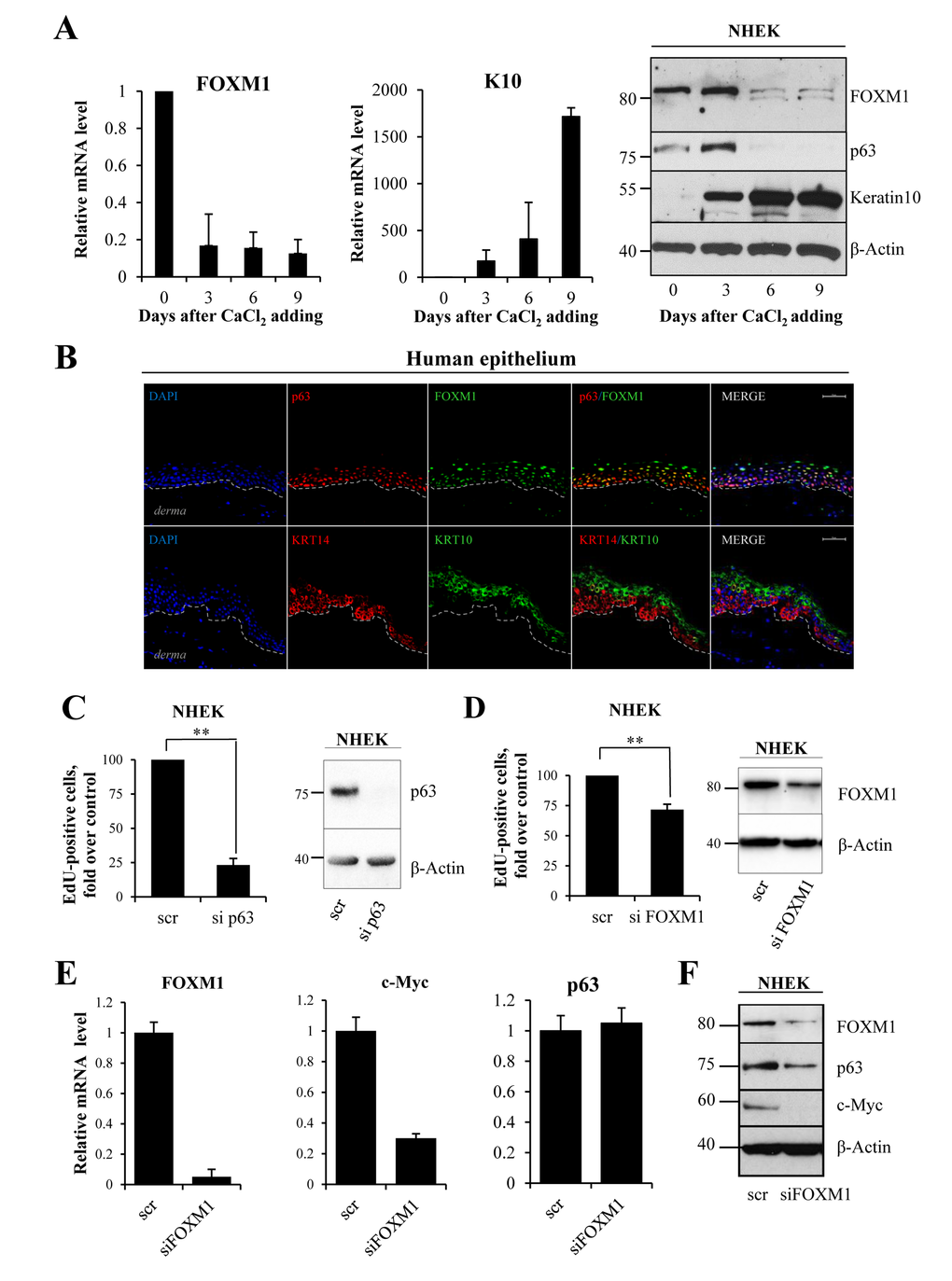
Figure 2.FOXM1 level correlates with proliferative status of keratinocytes(A) NHEKs were treated with calcium chloride to induce differentiation. FOXM1, Keratin10 and p63 levels were analyzed at 0, 3, 6 and 9 days after treatment by qRT-PCR and western blotting. Values reported are the average ± SD of four independent experiments. (B) Human epithelium was stained for FOXM1, p63 (lower layer marker), KRT10 (upper layers marker) and KRT14 (lower layer marker) and analyzed with confocal microscopy. DAPI was used for nucleus staining. The bar indicates 25 um. (C)NHEKs were silenced for p63 for 72 h, after which EdU-incorporation assays were performed. The percentage of EdU-positive cells was analyzed by FACS. (D)NHEKs were silenced for FOXM1 for 96 h, after which EdU-incorporation assay was performed. The percentage of EdU-positive cells was analyzed by FACS. Western blots confirm the silencing. Values reported are the average ± SD of two (for p63) and three (for FOXM1) independent experiments. **p-value <0.01 by Student's t-test. (E) Cells were silenced for FOXM1 for 96 h, after which the relative expression levels of FOXM1, c-MYC, and p63 were determined by qRT-PCR. (F)Western blot analysis of FOXM1, p63, and c-MYC levels.