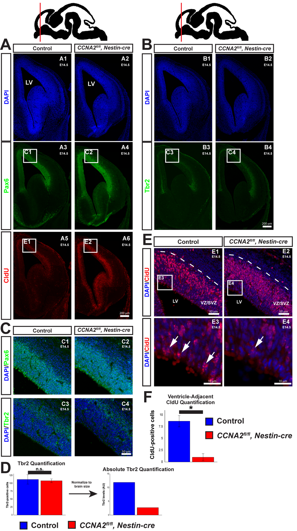
Figure 3.Delayed forebrain development in CCNA2fl/fl, Nestin-cre animals. (A) A timed-pregnant E14.5 dam was injected with CldU then euthanized. Embryos were stained for Pax6, CldU, and DAPI. Experimental condition is indicated above, molecular markers are color-coded on the left. LV = lateral ventricle. (B) Sections were stained for Tbr2. Experimental condition is indicated above, molecular markers are color-coded on the left. (C) Pax6 and Tbr2-positive cells are appropriately localized in CCNA2fl/fl, Nestin-cre forebrains, indicating preserved cytoarchitecture. Image locations are indicated in (A) and (B). (D) Quantification of Tbr2-positive cells. Cells were counted in a 100x180 μm counting frame in the VZ/SVZ. Tbr2-positive cells were unchanged within each counting frame. Accounting for the reduced size of the E14.5 VZ/SVZ there is a reduction in total Tbr2-positive cells. The y-axis is total Tbr2-positive cells. Unpaired t-test, n.s. = not significant, p > 0.05, n = 3 embryos per condition. (E) High magnification images of CldU staining in (A). Experimental condition is indicated above, molecular markers are color-coded on the left. Arrows indicate CldU-positive cells adjacent to the ventricle. (F) Quantification of CldU-positive cells adjacent to the lateral ventricle. CldU-positive cells were counted in a 100x10 μm bin adjacent to the ventricle. CldU-positive cells adjacent to the lateral ventricle were reduced in CCNA2fl/fl, Nestin-cre embryos. The y-axis is number of CldU-positive cells. Unpaired t-test, p < 0.05, n = 3 embryos per condition. Error bars for all graphs represent s.e.m.