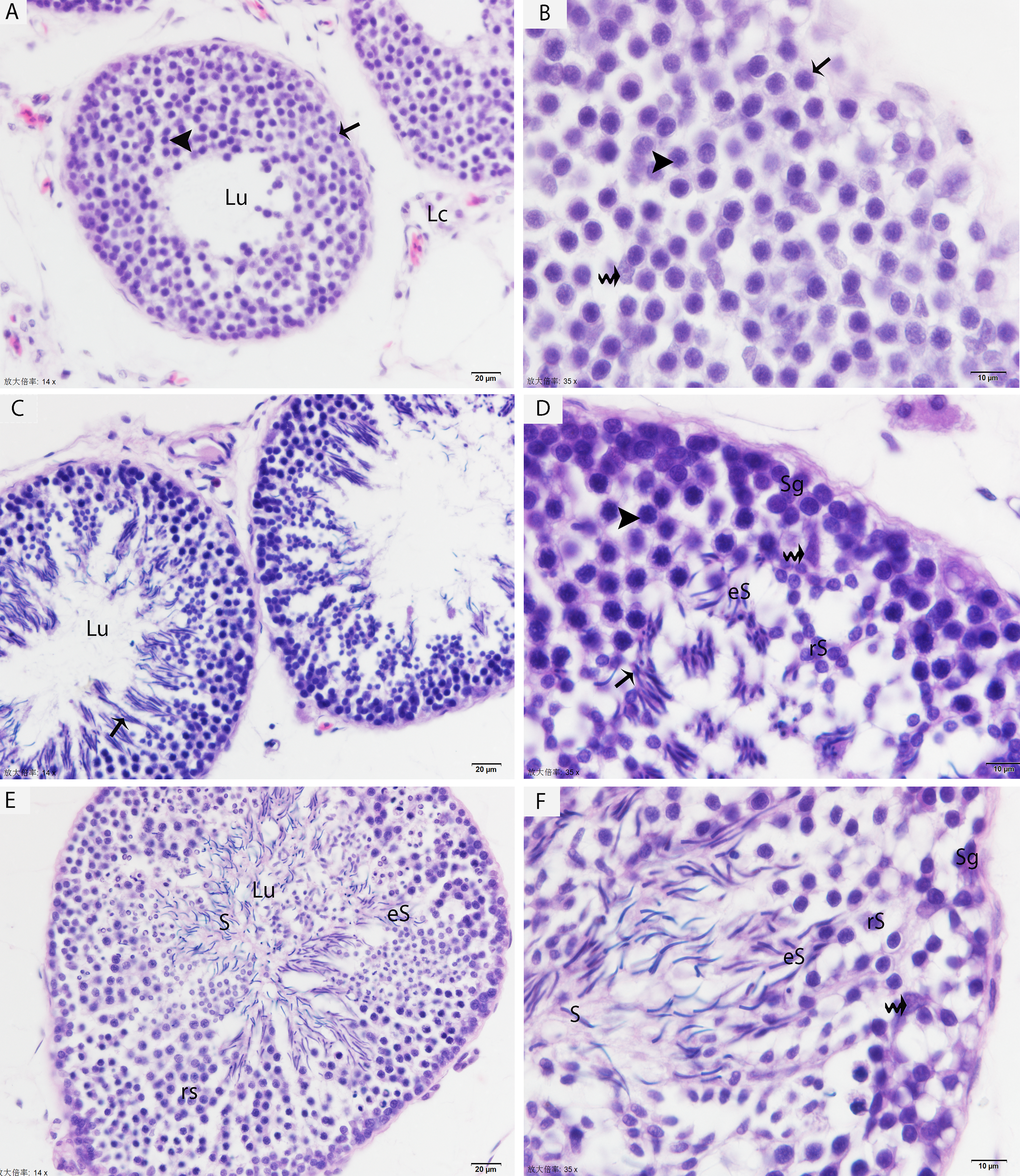
Figure 1.Light micrograph shows the histological structure of testis. (A, B) Seminiferous tubules show the spermatogonia (arrow) and few leptotene spermatocytes (arrowhead) in May. (C, D) Pachytene spermatocytes (arrowhead), round/elongated spermatids are present in the seminiferous epithelium, and spermatids are arranged in the sperm column (arrow) in July. (E, F) The round/elongated spermatids as well as free spermatozoa in the lumen and few spermatogonia are observed in October. LC: Leydig cell; Sg: spermatogonia; rS: round spermatid; eS: elongated spermatid; S: spermatozoa; Lu: lumen; (curved arrow): Sertoli cells. H & E stain. Scale bar= 20μm (A, C, E) and 10μm (B, D, F).