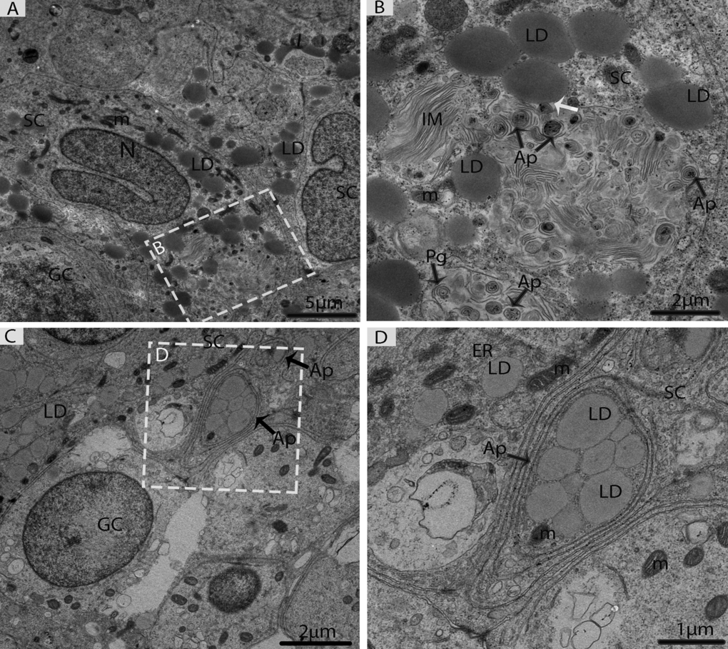
Figure 5.Electron micrograph of Sertoli cells during consumption of lipid droplets via lipophagy in May. (A) Sertoli cells contained numerous lipid droplets. (B) Illustration of Fig. A (rectangular area) shows the lipid droplets in contact with a phagophore and autophagosomes (white arrow). (C) Lipid droplets enclosed in the autophagosome. (D) Higher magnification of the square from Fig. C. SC: Sertoli cell; LD: lipid droplets; IM: isolation membranes; Ap: autophagosome; Pg: phagophore; GC: germ cell; m: mitochondria; ER: endoplasmic reticulum. Scale bar= 5μm (A), 2μm (B, C) and1μm (D).