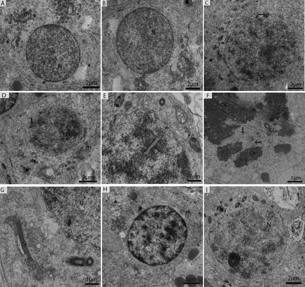
Figure 6.Electron micrograph of meiotic cells in July. (A) Primary spermatocytes in pre-leptotene stage. (B) Primary spermatocytes in leptotene. (C) Primary spermatocyte in zygotene beginning to form synaptonemal complexes (arrow). (D) Primary spermatocyte with visible synaptonemal complexes (arrow) in pachytene. (E) Synaptonemal complexes (arrow). (F) Thicker and dense chromosomes are attached with spindle fibers (arrow). (G) Golgi complex is observed close to the centrosome. (H) Primary spermatocytes in diplotene. (I) Secondary spermatocytes containing vacuolated mitochondria, including a few nuages. N: nucleus; m: mitochondria; Ch; chromosomes; CC: centrosome; Na: nuage; Gi: Golgi apparatus. Scale bar= 2μm (A, B, C, D, H, I) and 1μm (E, F, G).