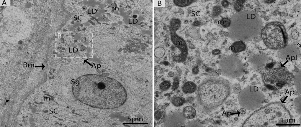
Figure 8.Electron micrograph of Sertoli cells in October. (A) Sertoli cells appeared with lipid droplets mitochondria and autophagosomes. (B) Illustration of panel (A) (rectangular area) clearly shows the mitochondria and autophagosomes attached to lipid droplets. SC: Sertoli cell; Sg: spermatogonia; Bm: basal membrane; Ap: autophagosome; Apl: autophagolysosome; LD: Lipid droplets; m: mitochondria. Scale bar= 5μm (A) and1μm (B).