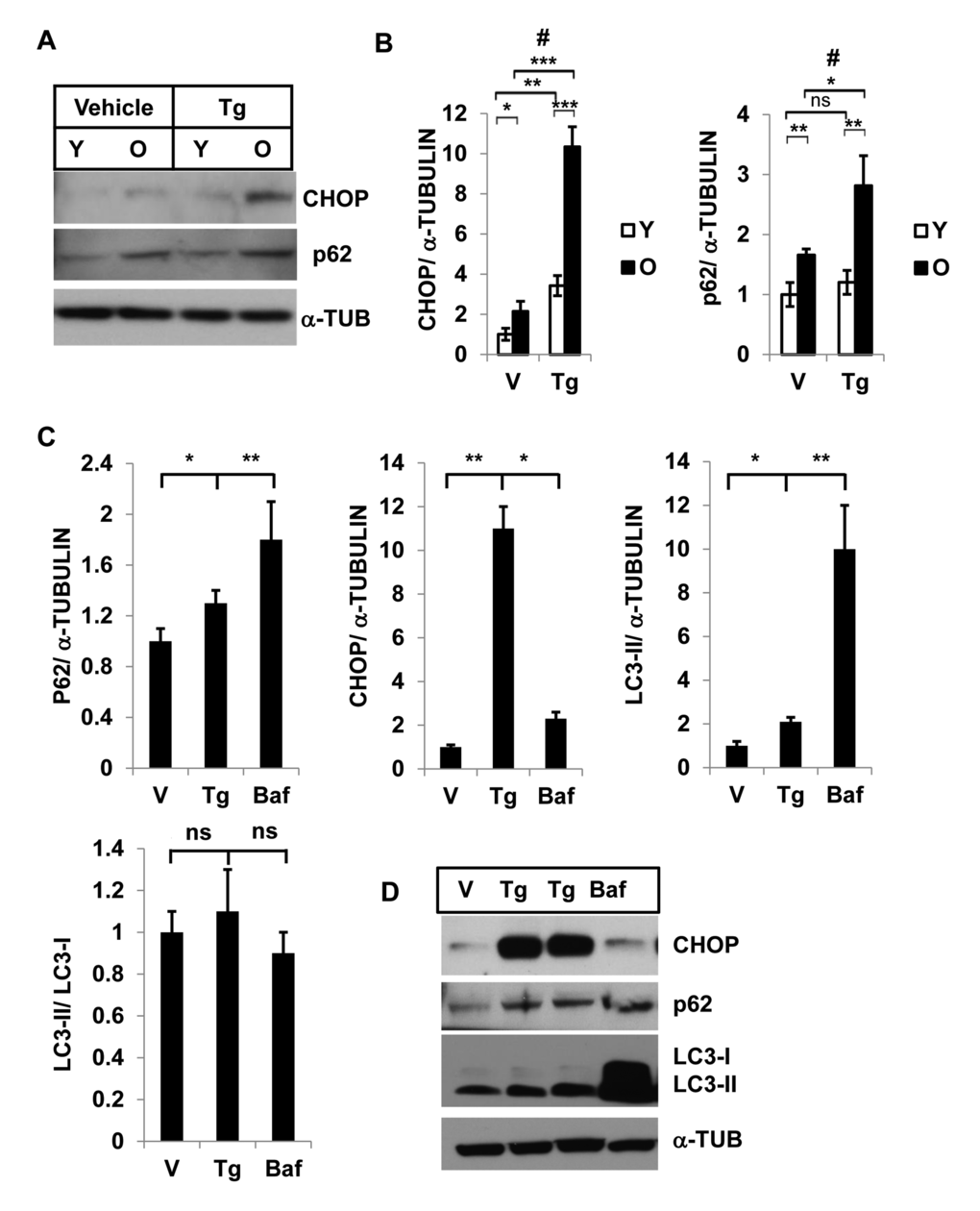
Figure 2.Interaction of autophagy and ER stress response in SVFs from adipose tissue and in 3T3-preadipocytes. Different levels of CHOP and SQSTM1/p62 in SVF lysates from young and old mice with vehicle or Tg treatments. (A) Western blot analysis of ER-stress response protein CHOP and autophagy associated protein SQSTM1/p62 in the SVF lysates from young (n=5) and old mice (n=3) treated with either vehicle (DMSO) or thapsigargin (30 nM) for 18h. The density of protein bands from three independent experiments were plotted in (B). Student’s t-test was performed using means and SD where *p<0.05 or **p< 0.01 and ***p<0.001 were designated significance levels. Symbol # indicated significance level (p<0.05) of two-way ANOVA analysis for the interaction between treatments (vehicle or Tg) and age factor (Y vs. O). (C, D) 3T3-preadipocytes were treated with either vehicle or Tg (30nM) or Baf (10nM) for 18 hrs and the protein levels of p62, CHOP and LC3-II were analyzed by western blots (D). The relative density of protein bands for P62, CHOP, LC3-II were plotted (C) after normalization with α-Tubulin. Student’s t-test was performed and *p<0.05 or **p <0.01 considered significant.