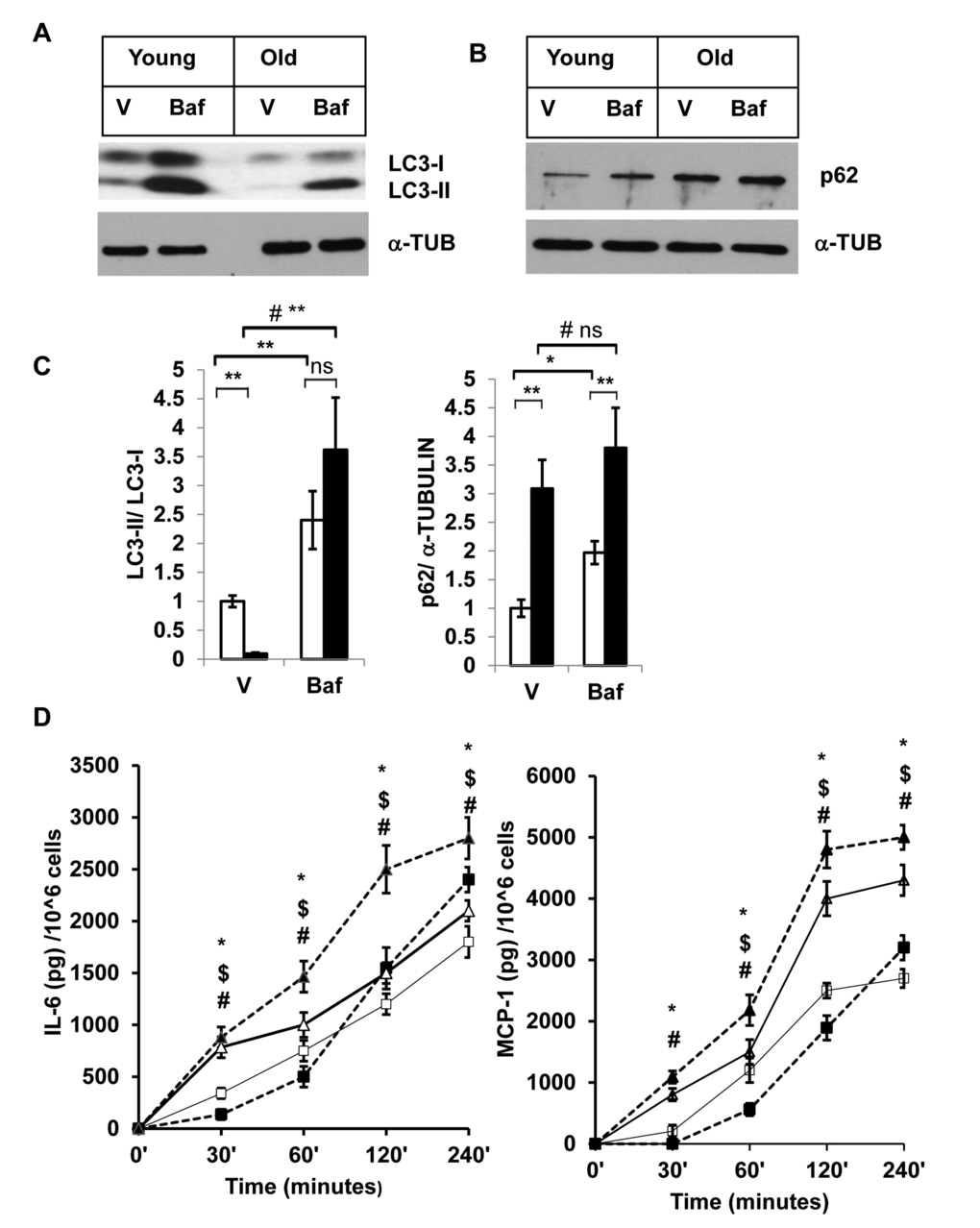
Figure 3.Enhanced accumulation of autophagy substrates in old SVFs and elevated pro-inflammatory cytokines release after autophagy block. (A and B) Western blot analysis of autophagy associated proteins SQSTM1/p62 and LC3-I and LC3-II in SVF lysates from young (n=5) and old mice (n=3) treated with either vehicle (DMSO) or Baf (10 nM) for 18h. The density of protein bands from three independent experiments were normalized with α-tubulin and plotted in (C). Student’s t-test was performed using means and SD where *p<0.05 or **p<0.01 was considered significant. Symbol # indicated significance level (p<0.05) of two-way ANOVA analysis for the interaction between treatments (vehicle or Tg) and age factor (Y vs. O). (D) Autophagy block results in elevated inflammatory cytokine production in old SVFs. Time dependent production of major pro-inflammatory cytokines (IL-6, MCP-1) by the SVF from young (square box) and old mice (triangle) treated with either vehicle (open) or Baf (filled) were analyzed by ELISA. Values were presented as mean + SD of three independent experiments. Significance of difference between means was determined by Student’s t-test and indicated by * p<0.05 for young and $ p<0.05 for old. Symbol # indicated the significance level (p<0.05) of two-way ANOVA analysis for the interaction between treatment (vehicle and Baf) and age factor (Y vs. O).