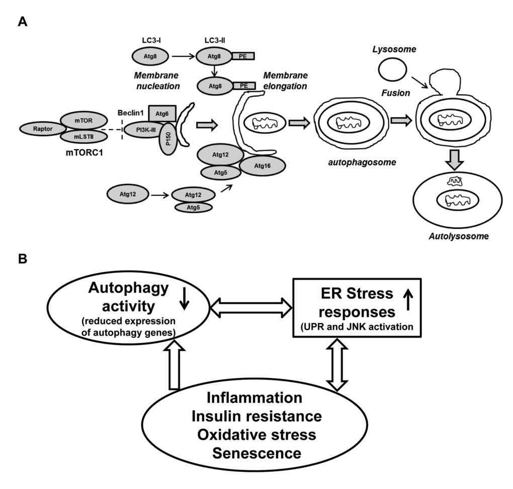
Figure 5.Schematic presentation of the autophagy process and its regulation: Connection with ER stress and oxidative stress and cellular senescence. (A) The formation of the initial membrane nucleation requires a kinase complex consisting of Beclin 1(Atg6), myristylated kinase (P150) and class III PI3K. The isolation membrane chooses its cargo (in this figure a mitochondria) and elongates until the edges fuse forming a double-membrane structure called an autophagosome. Two ubiquitin-like conjugation systems forming Atg8-PE (LC3-II) and Atg5-Atg12 are necessary for the elongation of the isolation membrane. The autophagosome matures by fusing with lysosomes, finally forming the autolysosomes. Abbreviations: mLST8: mammalian lethal with SEC13 protein 8, PI3KIII: phosphoinositidine 3-kinase class III, PE: phosphatidylethanolamine. (B) A simplified model of autophagy activity in aging SVFs: Diminished expression of autophagy machinery results in reduced autophagy activity. Compromised autophagy activity may lead to elevated ER stress, inflammation and oxidative damage resulting senescence. The senescent SVFs in turn further initiates a vicious cycle of compromised autophagy activity, elevated ER stress response and inflammation in the aging adipose tissue.