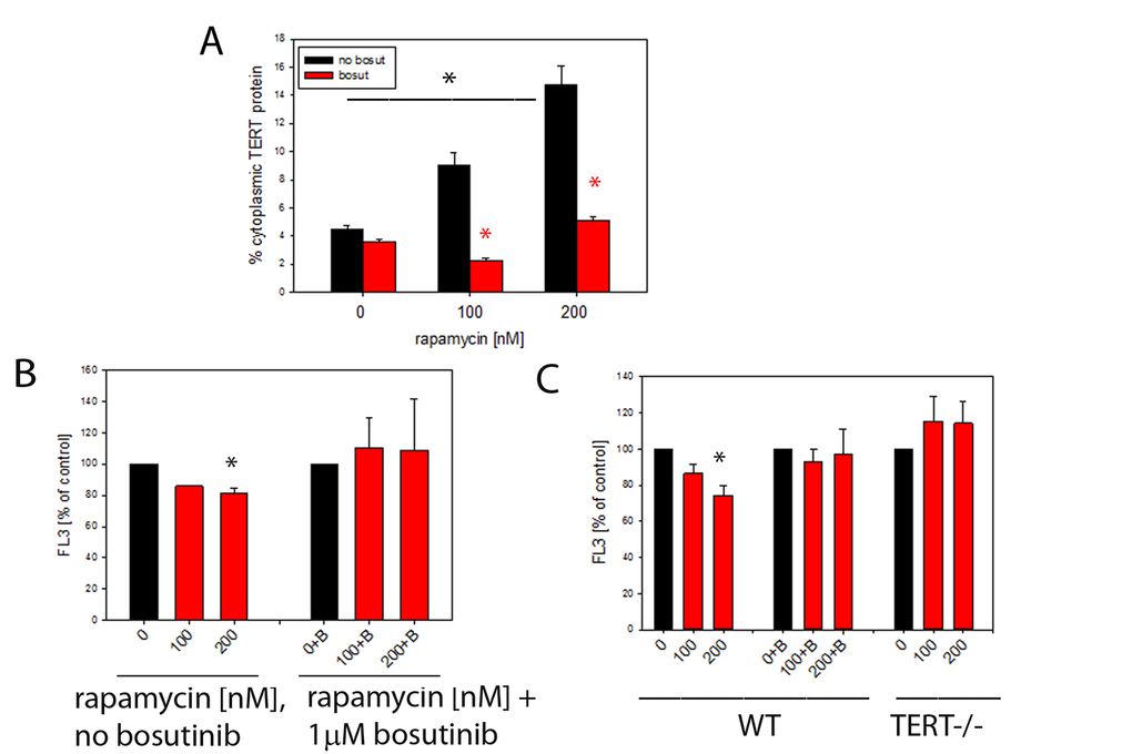
Figure 5.Rapamycin causes nuclear exclusion of TERT and reduces cellular ROS levels in a TERT-dependent fashion. (A) Extranuclear TERT levels (as fraction of total cellular anti-TERT immunofluorescence signal) in MCF-7 cells treated with the indicated concentrations of rapamycin. Bosutinib (1µM, red bars) was added to inhibit Src kinase. (*P<0.05, ANOVA for rapamycin concentration dependency and t-test for the effect of bosutinib at each rapamycin concentration) (B) Intracellular ROS levels (average DHR fluorescence intensity in flow cytometry, as percentage of untreated controls) in MCF-7 cells treated with rapamycin (concentrations indicated). B denotes additional treatment with 1 μM bosutinib. (* p<0.05, ANOVA, n=3-4 per condition) (C) Intracellular ROS levels in primary mouse ear fibroblasts from wild type (WT, left columns) and first generation TERT-/- (right columns) mice treated as in 5B. (2 way ANOVA, * p<0.05, for influence of rapamycin and genotype, n=7-8 per condition). Bosutinib treatment (middle column group) and lack of TERT (right columns) prevent decrease of ROS after rapamycin treatment in primary mouse ear fibroblasts, * p<0.05, One way ANOVA and Dunn’s post hoc test, n=7-8 per condition.