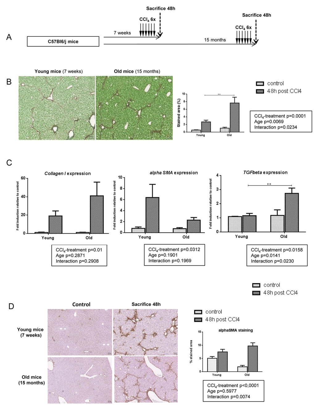
Figure 1.More severe liver fibrosis in old mice independently of profibrogenic processes. (A) CCl4 was injected three times a week for two weeks to young and old mice (n=6/group). Livers were harvested two days after the last injection. (B) Sirius red stained liver sections in CCl4-treated mice (magnification 80x). Scale bare 100µm. Collagen fibers were evaluated as percentage of stained area in the section (n=6/group). (C) Hepatic gene expression of Collagen I, alphaSma and Tgfbeta (Mean ± SEM) (n=6/group). (D) Activated stellate cells were identified by alphaSMA immunohistochemistry staining in young and old mice 48 hours after the last CCl4 injection (magnification 80x) (n=6/group). Scale bare 100µm. Statistical analysis was performed by two-way ANOVA for repeated measures (boxes) followed by Bonferroni’s post-hoc correction. **P<0.01 for differences between age groups.