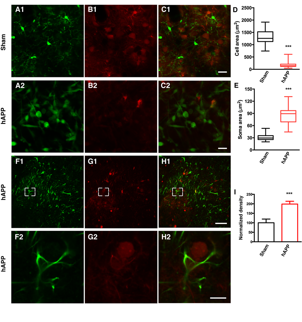
Figure 3.AAV-hAPP-FLAG induces microglial activation and astrocytosis.In vivo two-photon imaging of microglial activation in AAV-hAPP-SLA injected CX3CR1-GFP+/- mice. (A1-A2), GFP expressing microglial cells, (B1-B2), tdTomato and (C1-C2), merged. (1) Sham-operated mice indicating microglia cells in resting state, (2) AAV-hAPP-SLA injected mice indicating microglia cells in an activated state. Mean cell area (D) and mean soma area of microglia cells (E) in the sham and AAV-hAPP-SLA injected mice (Student’s test, P < 0.0001, n = 150 cells, 3 mice). (F-H) Representative images showing GFAP immunostaining in the PFC. Immunofluorescence images at low (1) and high (2) magnification for GFAP (F), Aβ oligomers (G) and merged (H). (I) Quantification of GFAP density. The density was normalized to control levels. The error bar is ± SEM. (Student’s test, P < 0.001, 6 slices were analyzed from 3 control mice and 6 slices from 3 AAV-hAPP-SLA injected mice). Scale bars = 50 µm, for H2 scale bar = 10 µm.