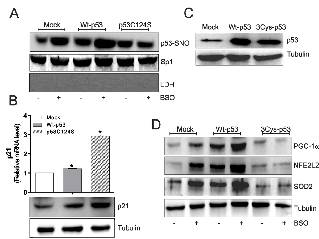
Figure 2.p53C124S mutant does not undergo S-nitrosylation after BSO treatment in C2C12 myoblasts. (A) C2C12 myoblasts were transfected with pcDNA3.1 vector containing cDNA for wild type p53 (Wt-p53), single p53 mutant in DBD (p53C124S) or with empty vector (Mock). After 15 h from transfection myoblasts were treated with 1 mM BSO for 24 h. Nuclear proteins (500 μg) were subject to S-NO derivatization with biotin. After Western blot the nitrocellulose was incubated with p53 antibody for detection of p53-SNO. Sp1 was used as loading control. The possible presence of cytoplasmic contaminants was tested by incubating nitrocellulose with rabbit anti-LDH. (B) Upper: Total RNA was isolated and relative mRNA level of p21 was analyzed by RT-qPCR . Data are expressed as means ± S.D. (n=3; *p<0.05). Bottom: Cells were lysed and 20 μg of proteins were loaded for Western blot analysis of p21. Tubulin was used as loading control. (C, D) C2C12 myoblasts were transfected with pcDNA3.1 vector containing cDNA for wild type p53 (Wt-p53), triple p53 mutant in DBD (C277S, C275S and C124S) (3Cys-p53) or with empty vector (Mock). After 15 h from transfection myoblasts were treated with 1 mM BSO for 24 h. Cells were lysed and 20 μg of proteins were loaded for Western blot analysis of p53, PGC-1α, NFE2L2 and SOD2. Tubulin was used as loading control. All the immunoblots reported are from one experiment representative of five that gave similar results.